Warning: mkdir(): Permission denied in /home/virtual/lib/view_data.php on line 81

Warning: fopen(upload/ip_log/ip_log_2024-04.txt): failed to open stream: No such file or directory in /home/virtual/lib/view_data.php on line 83

Warning: fwrite() expects parameter 1 to be resource, boolean given in /home/virtual/lib/view_data.php on line 84
Histochemical studies on the relationship between the ascarides of man and pig.
| Home | E-Submission | Sitemap | Contact us |  
top_img
Korean J Parasito Search

CLOSE

Korean J Parasito > Volume 1(1):1963 > Article

Original Article
Korean J Parasitol. 1963 Jun;1(1):15-21. English.
Published online Mar 20, 1994.  http://dx.doi.org/10.3347/kjp.1963.1.1.15
Copyright © 1963 by The Korean Society for Parasitology
Histochemical studies on the relationship between the ascarides of man and pig.
Jung-Kyun Chu and Hyung-Sup Lee
Department of Parasitology, Pusan National University, Medical School, Korea.
Department of Biochemistry, Pusan University, Medical School, Korea.
Abstract

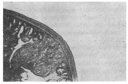
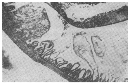
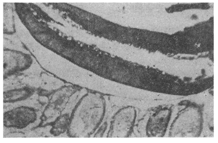
1. The histochemical study, especially the demonstration of alkaline phosphatase and acid phosphatase was carried out in order to differentiate ascarides of human and pigs.

2. The experimental material were obtained from naturally contaminated men and pigs. As the histochemical staining methods the Gomori's was applied for acid phosphatase and Takeuchi and Takami's for alkaline phosphatase.

3. The results obtained were summerized as follows

a. In the pig's ascarides, alkaline phosphatase was richly found in the subcuticular tissue, lateral line, median line, strial zone and epithelial cells of the intestine, epithelial cell and basal membrane of the ovary, the same part of the uterus and also in eggs.

Acid phosphatase in the pig's ascarides were distributed in the same part as alkaline phosphatase. It, however, was darker brown in the soft tissue of the lateral line, epithelium of excretory canal, median bundle, whole zone of the intestine and intestinal contents.

b. In the human ascarides, the alkaline phosphatase was distributed in the testes and the parts where the acid phosphatase was found in the pig ascarides.

The acid phosphatase in the human ascarides was demonstrated in the subcuticular tissue, soft tissue of lateral line, epithelium of excretory cells, strial zone, transparent zone, granular zone and epithelial zone of esophagus and intestine, ovary, ova in the uterus, epithelial cell and basal membrane of the uterus and in testes.

4. In the pig's ascarides, the area of distribution of alkaline phosphatase was restricted, but that of acid phosphatase was wider.

5. In human ascarides, the area of distribution of alkaline phosphatase and acid phosphatase was not significantly different, but in some part showed slight difference.

6. Above mentioned finding suggest that the distribution of phosphatase could be utilized for the differentiation of ascarides of human and pig.

Figures


Fig. 1
Distribution of the carbonic anhydrase in the wall of Ascaris suum.


Fig. 2
Distribution of the alkaline phosphatase in the wall and midgut of Ascaris lumbricoides.


Fig. 3
Distribution of the alkaline phosphatase in the midgut of Ascaris lumbricoides.

References
1. Gomori G. Proc Soc Exp Biolet Med 1939;42:23.
2. Gomori G. Arch Path 1941;23:189.
3. Roger's WP. Nature 1947;195:374.
4. Bullock WL. J Morph 1947;84:185.
 
5. Bullock WL, et al. J Parasit 1950;36 Suppl:30.
6. Bullock WL. Exp Parasit 1953;2:152–162.
Editorial Office
Department of Molecular Parasitology, Samsung Medical Center, School of Medicine, Sungkyunkwan University,
2066 Seobu-ro, Jangan-gu, Suwon 16419, Gyeonggi-do, Korea.
Tel: +82-31-299-6251   FAX: +82-1-299-6269   E-mail: kjp.editor@gmail.com
About |  Browse Articles |  Current Issue |  For Authors and Reviewers
Copyright © 2024 by The Korean Society for Parasitology and Tropical Medicine.     Developed in M2PI