Warning: mkdir(): Permission denied in /home/virtual/lib/view_data.php on line 81
Warning: fopen(upload/ip_log/ip_log_2024-04.txt): failed to open stream: No such file or directory in /home/virtual/lib/view_data.php on line 83
Warning: fwrite() expects parameter 1 to be resource, boolean given in /home/virtual/lib/view_data.php on line 84 A study on the chemotherapy in clonorchiasis. Report 3. The patho-histological study on the liver of rabbit healed from clonorchiasis by chemotherapy
A study on the chemotherapy in clonorchiasis. Report 3. The patho-histological study on the liver of rabbit healed from clonorchiasis by chemotherapy
Suck Yong Kang, M.D.,Yong Soo Chun, M.D.,In Kyun Loh, M.D. and Eui Keun Ham, M.D.
Department of Internal Medicine and Institute of Endemic Diseases, College of Medicine, Seoul National University, Seoul, Korea.
School of Public Health, Seoul National University, Korea.
Department of Pathology, College of Medicine, Seoul National University, Korea.
Abstract
In the experimental rabbits which were previously infected with Clonorchis sinensis and were thereafter administered with Dithiazanine iodide, the patho-histological and some histochemical changes were observed in the healed liver(76 days after stoppage of egg excretion). And also, the patho-histological changes which appeared in the infected and healed livers respectively were compared from each other.
The following results and conclusion were obtained.
1) In the infected group, grossly the liver showed a considerable increase of its weight, an increased vascular marking of its surfaces and an increase in consistency and thickness of the bile ducts. However, the healed group grossly showed no remarkable changes in the liver.
2) In the infected group, microscopically the liver showed a marked adenomatous hyperplasia of bile ducts, proliferation of bile ductules and marked periductal and periductular fibrosis with infiltration of chronic inflammatory cells in portal spaces. A histochemical study showed that the increased fibrous tissue consists largely with collagenous fibers and partly with reticulum fibers.
3) In the group with healed liver microscopically the liver showed much improvement in the histopathological changes compared to those of the infected group. In the healed lover there remained only slight histopathological changes mainly in portal spaces of the liver, such as considerably diminished periductal and periductular fibrosis. The slightly increased fibrous tissue consists largely with reticulum fibers and partly with collagen fibers.
Figures
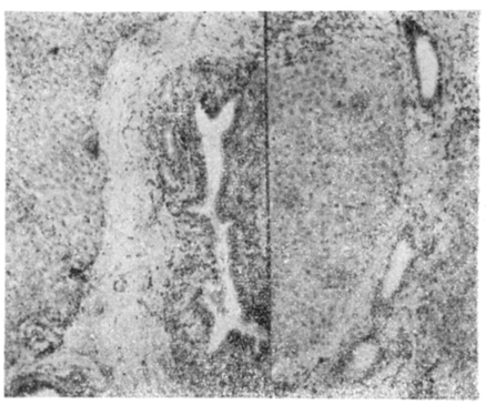
Fig. 1 Liver showing marked adenomatous hyperplasia of bile duct(Rt) and bile ductule proliferation (Lt), No.2 of C. sinensis infected, H-E stain. ×100.
Fig. 2 Liver showing relatively well preserved bile ductule (Lt) and bile ductules (Rt), No.7 of completely healed from clonorchiasis, H-E stain. ×100.
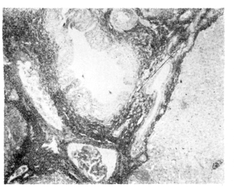
Fig. 3 C. sinensis worm in adenomatous proliferated bile duct and surrounding dense collagenous fiber band, No. 2 of C. sinensis infected, Vangieson stain, ×100.
Fig. 4 Liver showing well preserved bile duct with normal degree of collagenous fiber bland, No.7 of completely healed from clonorchiasis, Vangieson stain, ×100.
Fig. 5 Liver showing adenomatous hyperplasia of bile duct and surrounding dense fibrosis with scanty reticulum fiber, No.2 of C. sinensis infected, Reticulum silver stain, ×100.
Fig. 6 Liver showing well preserved bile duct (Lt) and ductules (Rt) with surrounding rather prominent amounts of reticulum fiber, No. 7 of completely healed from clonorchiasis, Reticulum silver stain, ×100.
Kang SY, Loh IK, Chun YS, Lim DS. [A Study On The Chemotherapy In Clonorchiasis: Report 1. An Experimental Study On Chemotherapy With Dithiazanine Iodide And Bithionol Sulfoxide In Clonorchiasis]. Korean J Parasitol 1965;3(1):19–30.
2.
Yamaguchi T, et al. Jap J Parasit 1961;10(6):697–704.
3.
Yokogawa M, et al. Jap J Parasit 1965;14(3):233–242.
4.
Yokogawa M, et al. Jap J Parasit 1965;14(6):526–533.