Warning: mkdir(): Permission denied in /home/virtual/lib/view_data.php on line 81
Warning: fopen(upload/ip_log/ip_log_2024-04.txt): failed to open stream: No such file or directory in /home/virtual/lib/view_data.php on line 83
Warning: fwrite() expects parameter 1 to be resource, boolean given in /home/virtual/lib/view_data.php on line 84 Study on Metagonimus yokogawai(Katsurada, 1912) in Korea I. On the metacercaria, its distribution in the second intermediate host and the development in the final host
Study on Metagonimus yokogawai(Katsurada, 1912) in Korea I. On the metacercaria, its distribution in the second intermediate host and the development in the final host
Byong Seol Seo and Nam Tae Hong
Department of Parasitology and Institute of Endemic Diseases, College of Medicine, Seoul National University, Korea.
Abstract
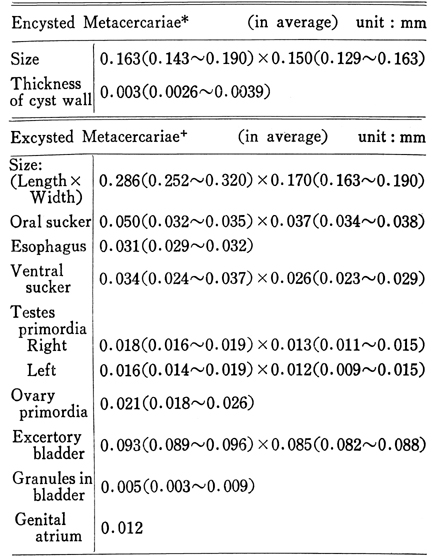
1) The metacercariae of Metagonimus yokogawai were isolated from the sweetfish Plecoglossus altivelis, clollected at Hwagae, South Kyongsang Do, one of the newly known endemic foci of metagonimiasis in Korea. The body structure of metacercaria of M.yokogawai was described and the measurements of the excysted metacercaria were also made.
2) In order to know the distribution of metacercariae within the host, the rate of infection and the intensity of infection, a total of 10 sweet fishes was examined and it was found all infected, from which a total of 38,511 metacercariae was isolated. The number of metacercariae in a fish varied from 219 to 14,427. The average number of metacercaria per fish was 3,851.
The distribution of metacercariae in the four divided parts of fish was observed in the following order; number of metacercaria in the muscles; 2,417 (62.8%), in the subcutaneous tissues; 1,126.9 (29.3%), and on the scale; 291.9(7.7%).
3. The development of the metacercaria of M. yokogawai in the mouse host was experimentally traced every day for 10 days after infection . In an earlier period of infection, the growth rate of the genital primordia was distinctly high, particularly in the testes. The seminal receptable and seminal vesicle became clearly recognized in measurable size at 4 days after infection. The vitelline follicles and their ducts were also first visible in the living specimens at 6 days after infection. The oral sucker was larger in size than the ventral sucker in an early stage of the worms, however after 7 days after infection it reversed. The posterior part of body began to extend since two days after infection. Fully matured worms were able to collect only after 7 days after infection. At this stage, the body of worm became 0.7 mm long and 0.3 mm wide. The first positive appearance of eggs in the uterine tubule and in feces was on the 6th day and 10th day of infection, respectively.
In an earlier stage of infection, the worms were found mostly in the upper portion of small intestine and the recovery rates of the worms were high, however according to the course of infection in later stage they were seen rather in the lower part of the intestine and the recovery rate also decreased.
Figures
Fig. 1 Metacercaria of MMetagonimus yokogawai ventrally bent for revolving movement.
Fig. 2 Distribution of metacercariae of M. yokogawai in the sweetfish, Plecoglossus altivelis.
Fig. 3 Distribution of worms in the intestine and recovery rate by various intervals after infection.
1-5: parts of the small intestine, 6: large intestine
Plate I Fig. 1 and 2: Metacercaria of M. yokogawai.
Table 1 Measurements of metacercaria of M. yokogawai
Table 2 Distribution of metacercariae of Metagonimus yokogawai in Plecoglossus altivelis
Table 3 Measurements(in average) of Developing Worms in Mouse Host carrying Age of Infection
References
1.
Choi DW, Lee JT, Hwang HK, Shin YD. [Studies Of The Larval Trematodes From Brackish Water Fishes: 2. Observation On Metagonimus Yokogawai Katsurada, 1912]. Korean J Parasitol 1966;4(1):33–37.
2.
Chun SK. Bulletin of Pusan Fisheries College 1960;3(1,2):31–39.
3.
Chun SK. Bulletin of Pusan Fisheries College 1960;3(1,2):24–30.
4.
Furuyama T. J Chosen Med Ass 1930;20:251–252.
5.
Ito J, et al. Jpn J Parasit 1957;6(3,4):356.
6.
Kang SY, et al. J Korean Med Ass 1964;7(5):470–476.
7.
Komiya Y, et al. Jpn J Parasit 1958;7(1):1, 11.
8.
Lee JT. [Studies on the metacercariae from fresh water fishes in the Kum-Ho River]. Korean J Parasitol 1968;6(3):77–99.
9.
Oshima T, et al. Jpn J Parasit 1966;15(1):161–168.
10.
Seo BS, Rim HJ, Loh IK, Lee SH, Cho SY, Park SC, Bae JW, Kim JH, Lee JS, Koo BY, Kim KS. [Study On The Status Of Helminthic Infections In Koreans]. Korean J Parasitol 1969;7(1):53–70.
11.
Yokogawa M, et al. Jpn J Parasit 1968;17(6):540–545.