Warning: mkdir(): Permission denied in /home/virtual/lib/view_data.php on line 81
Warning: fopen(upload/ip_log/ip_log_2024-04.txt): failed to open stream: No such file or directory in /home/virtual/lib/view_data.php on line 83
Warning: fwrite() expects parameter 1 to be resource, boolean given in /home/virtual/lib/view_data.php on line 84 An imported case of Kala-azar in Korea
Je Geun Chi,Young Kee Shong,Sung Tae Hong,*Soon Hyung Lee,*Byong Seol Seo,* and Kwang Won Choe**
Department of Pathology, College of medicine, Seoul National University, Korea.
*Department of Parasitology, College of medicine, Seoul National University, Korea.
**Department of Internal Medicine, College of medicine, Seoul National University, Korea.
Abstract
An imported case of Kala-azar in a 26-year-old Korean man is reported. The diagnosis was made by liver needle biopsy. Amastigotes were seen in Kupffer cells under light microscope, and their characteristic ultrastructural features were recognized under the electron microscope.
This case represents an imported disease from Saudi Arabia where the patient spent one year as a construction worker, 8 months before the onset of the disease. This report also signifies the second description of Kala-azar in Korea, and the first case of Kala-azar imported from Saudi Arabia.
This patient was successfully treated with sodium antimony gluconate (Pentostam), and follow up liver biopsy showed focal fibrous scar and otherwise normal liver without demonstrable organism.
Figures
Fig Temperature curve before and after Pentostam treatment.
Figs. 1-4 Fig. 1. Photograph of the patient's abdomen to show hepatosplenomegaly. The lower margins of the liver and the spleen are outlined with black ink.
Fig. 2. Low power view of the first liver biopsy before treatment. Note patchy area of necrosis and disorganization of lobular architecture in the right half. H&E ×100.
Fig. 3. High power view of amastigote in Kupffer cells of the junction of necrotic zone and relatively preserved area. H&E ×1,000.
Fig. 4. Liver biopsy picture after treatment showing foci of dense fibrosis and otherwise well preserved liver tissue. No organism is demonstrated. H&E ×100.
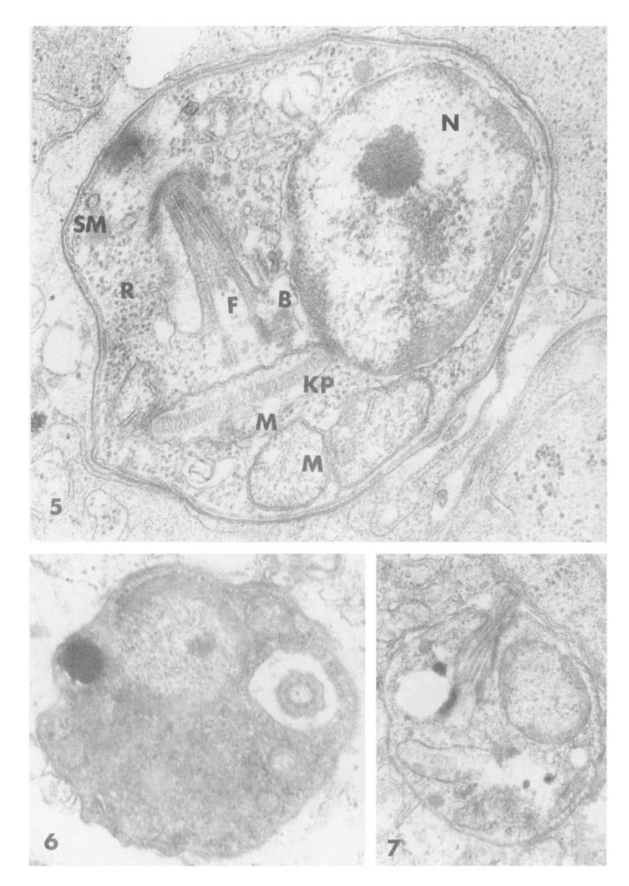
Figs. 5-7 Fig. 5. TEM picture of typical leishmania (amastigote) ×47,600, abbreviations as follows; N: nucleus with a central karyosome and peripheral chromatin, SM: subpellicular microtubule, M: mitochondria, F: flagellum, KP: kinetoplast, B: basal body, R: ribosomes.