Warning: mkdir(): Permission denied in /home/virtual/lib/view_data.php on line 81
Warning: fopen(upload/ip_log/ip_log_2024-04.txt): failed to open stream: No such file or directory in /home/virtual/lib/view_data.php on line 83
Warning: fwrite() expects parameter 1 to be resource, boolean given in /home/virtual/lib/view_data.php on line 84 A study on the fine structure of Clonorchus sinensis, a liver Fluke V. The mature spermatozoa
A study on the fine structure of Clonorchus sinensis, a liver Fluke V. The mature spermatozoa
Kye Heon Jeong and Han Jong Rim
Department of Biology, Soonchunhyang University, Onyang, Asan, Chungnam, Korea.
Department of Parasitology and Institute for Tropical Endemic Diseases, College of Medicine, Korea University, Korea.
Abstract
An ultrastructural study on the mature spermatozoa of Clonorchis sinensis was carried out. For this study, the liver flukes were collected from the livers of rabbits and rats artificially infected with the metacercariae obtained from the fresh water fish, Pseudorasbora parva. Six-month old worms were used. The collected liver flukes were washed with 0.85 percent saline solution and then immediately moved to cold 2 percent glutaraldehyde buffered with 0.l M Millonig's phosphate buffer (pH 7.4). The materials were dissected into appropriate pieces in the fixative about 30 minutes after beginning of the fixation. Two hours later the materials containing the seminal receptacle were rinsed several times with the buffer and were secondarily fixed with cold, buffered 1 percent osmium tetroxide for 2 hours. The fully fixed tissue blocks were dehydrated in a series of graded concentrations of acetone and were embedded in Epon 812 mixture. Thin sections obtained from LKB-5 ultramicrotome were stained with uranyl acetate and Reynold's lead citrate. Observations of the sections were carried out with JEM-100CX II electron microscope. In general, the mature sperm was long thread-like form with a sickle-shaped head. According to the longitudinal sectioned view of the sperm tail, the nucleus seemed to be spirally coiled and run a little far along the tail. The acrosome was not observed. The cytoplasm of the tail was biflagellated as usual in trematodes. Unlike other platyhelminth spermatozoa, the sperm tail of Clonorchis sinensis showed the [9+2] pattern in the microtubular arrangement. The mitochondria with poorly developed cristae were observed throughout the middle piece. The middle piece of the tail showed dull ladder or triangular shapes with the two flagella at the bottom. But, the principal piece of the tail was slightly flattened cylindrical shape with two flagella within the cytoplasm. The end piece was uniflagellated. It was not clearly identified whether the end piece was subdivided into two by flagellum or the lengths of the two flagella were different. The glycogen granules were rich in the cytoplasm throughout the length of the spermatozoa. These granules might be the energy source for the movement of the spermatozoa.
Figures
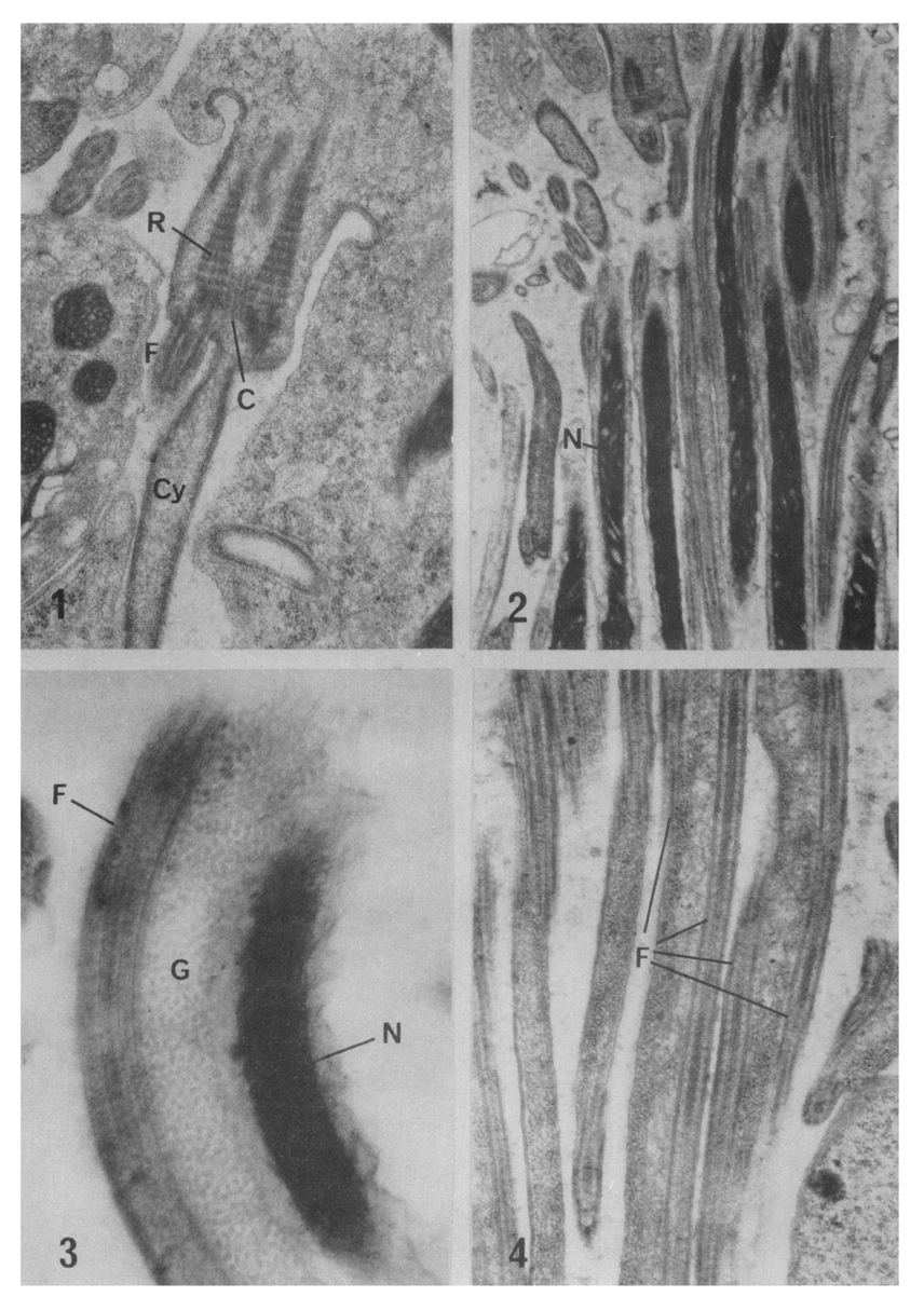
Figs. 1-4 Fig. 1. A spermatid undergoing spermiogenesis. The rootlets (R) serve as sites of origin of the flagella (F). Centriole (C) is located between the rootlets. The cytoplasm (Cy) is getting elongation to form the tail with the flagella. ×25,700.
Fig. 2. Longitudinal view of the anterior part of the sperm tail. The spirally coiled nucleus (N) rums along the flagella. ×12,900.
Fig. 3. High magnification of the anterior part of the sperm tail. The cytoplasm contains rich glycogen gramules (G). ×55.100.
Fig. 4. Longitudinal view of the middle piece of the tail. The cytoplasm of the tail is biflagellated (F). ×23,600.
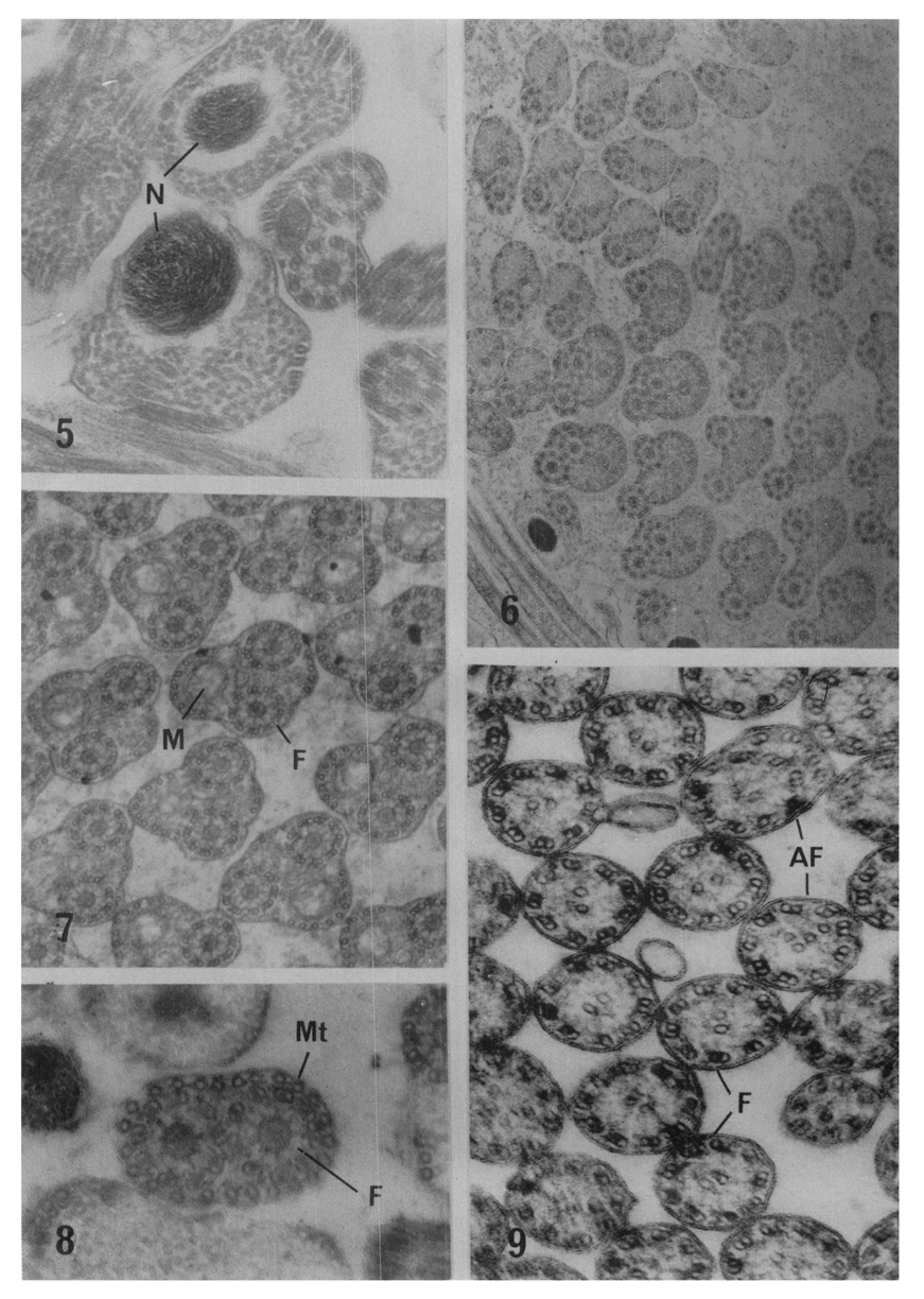
Figs. 5-9 Fig. 5. The head part of the sperm with dense nucleus (N). ×53,600.
Fig. 6. Cross sectioned view of the anterior middle pieces of the sperms with two flagella. ×29,200.
Fig. 7. Cross sectioned view of the middle pieces. Mitochondria (M) with cristae are seen with two flagella(F). ×43,700.
Fig. 8. Cross sectioned view of the principal piece. The two flagella are closely neighbored and surrounded by the cortical microtubules (Mt) sited right inside the cell membrane. ×87,900.
Fig. 9. Cross sectioned view of the end pieces The [9+2] arrangement of microtubules in the flagella (F) are seen. A few abnormal flagella (AF) are also observed.
References
1.
Anderson WA, Personne P. The localization of glycogen in the spermatozoa of various invertebrate and vertebrate species. J Cell Biol 1970;44(1):29–51.
2.
von Bonsdorff CH, Telkkä A. The spermatozoon flagella in Diphyllobothrium latum (fish tapeworm). Z Zellforsch Mikrosk Anat 1965;66(5):643–648.
3.
Burton PR. Gametogenesis and fertilization in the frog lung fluke, Haematoloechus medioplexus Stafford (Trematoda: Plagiorchiidae). J Morphol 1960;107:93–121.
4.
Burton PR. Fine structure of the reproductive system of a frog lung fluke. II. Penetration of the ovum by a spermatozoon. J Parasitol 1967;53(5):994–999.
5.
Burton PR. Effects of various treatments on microtubules and axial units of lung-fluke spermatozoa. Z Zellforsch Mikrosk Anat 1968;87(2):226–248.
6.
Burton PR. Fine structure of the reproductive system of a frog lung fluke. 3. The spermatozoon and its differentiation. J Parasitol 1972;58(1):68–83.
7.
Fujino T, et al. Japanes J Parasit 1977;26(4):240–255.
8.
Grant WC, Harkema R, Muse KE. Ultrastructure of Pharyngostomoides procyonis Harkema 1942 (Diplostomatidae). I. Observations on the male reproductive system. J Parasitol 1976;62(1):39–49.
9.
Gresson RAR. Parasitol 1965;55:117–125.
10.
Gresson RA, Perry MM. Electron microscope studies of spermateleosis in Fasciola hepatica L. Exp Cell Res 1961;22:1–8.
11.
Hendelberg J. Zool Bidr Uppsala 1962;35:569–587.
12.
Hendelberg J. Zool Bidr Uppsala 1969;38:1–50.
13.
Jeong KH, Rim HJ, Yang HY, Kim WK, Kim CW. A Morphological Study On Spermatogenesis In The Liver Fluke, Clonorchis Sinensis. Korean J Parasitol 1976;14(2):123–132.
14.
Kitajima EW, Paraense WL, Correa LR. The fine structure of Schistosoma mansoni sperm (Trematoda: Digenea). J Parasitol 1976;62(2):215–221.
15.
Lumsden RD. Microtubules in the peripheral cytoplasm of cestode spermatozoa. J Parasitol 1965;51(6):929–931.
16.
Nez MM, Short RB. Gametogenesis in Schistosomatium douthitti (Cort) (Schistosomatidae: Trematoda). J Parasitol 1957;43(2):167–182.
17.
Sato M, Oh M, Sakoda K. Electron microscopic study of spermatogenesis in the lung fluke (Paragonimus miyazakii). Z Zellforsch Mikrosk Anat 1967;77(2):232–243.
18.
Silveira M, et al. Protoplasm 1964;59:240–265.
19.
Tulloch GS, Hershenov BR. Fine structure of platyhelminth sperm tails. Nature 1967;213(5073):299–300.