Warning: mkdir(): Permission denied in /home/virtual/lib/view_data.php on line 81
Warning: fopen(upload/ip_log/ip_log_2024-04.txt): failed to open stream: No such file or directory in /home/virtual/lib/view_data.php on line 83
Warning: fwrite() expects parameter 1 to be resource, boolean given in /home/virtual/lib/view_data.php on line 84 Tegumental ultrastructures of Echinostoma hortense observed by scanning electron microscopy
Tegumental ultrastructures of Echinostoma hortense observed by scanning electron microscopy
Soon Hyung Lee,Sung Jong Hong,Jong Yil Chai,Sung Tae Hong and Byong Seol Seo
Department of Parasitology and Institute of Endemic Diseases, College of Medicine, Seoul National University, Seoul 110, Korea.
Abstract
The tegumental ultrastructures of Echinostoma hortense adults were observed by scanning electron microscopy. The worms of 4 weeks of age were harvested from albino rats experimentally infected with the metacercariae obtained from the loach. The results were as follows: The worms were leaf-like and their anterior end portion, including oral sucker and head crown, ventrally curved to face posteriorly. The tegument of whole body was wrinkled transversely and covered with cobblestone-like cytoplasmic processes. The oral sucker had roundly swollen (type II) sensory papillae on the ventral half of its lip and uni-ciliated knob-like (type I) sensory papillae, arranged in 2-3 rows, on the dorsal outer surface. Aspinous ventral sucker had many of type I papillae arranged in a circular band on its outer surface. The tegument around the genital opening was of similar feature to the ventral sucker, but sensory papillae were hardly found around the former. Scale-like spines with broad base and round tip were distributed densely on the tegument anterior to the ventral sucker but they became sparse in posterior half of the ventral surface, finally to disappear at posterior extremity. A few number of type I papillae were observed on the ventral surface. The results suggest that the tegument of E. hortense is similar to that of other echinostomes especially E. revolutum. But the number and arrangement of collar spines, and/or the type and distribution of sensory papillae seem characteristic features of E. hortense differed from other echinostomes.
Figures
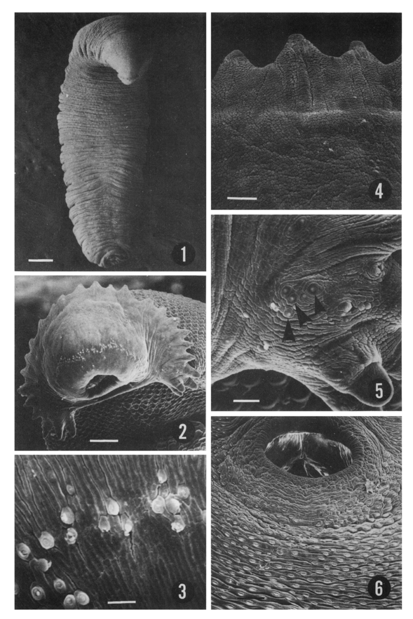
Figs. 1-6 Fig. 1. Ventral view of 4-week old E. hortense. Bar=300µm.
Fig. 2. Its head part showing oral sucker, head crown, and 27 collar spines including end group ones. Note the arrangement of sensory papillae on the lip of oral sucker. Bar=60µm.
Fig. 3. Ciliated knob-like papillae (type I) aligned in 2~3 rows in the manner of "zigzag" on the outer surface of oral sucker. Bar=3µm.
Fig. 4. Dorso-median part of head crown showing several collar spines covered with square-shaped or rectangular cytoplasmic processes. Bar=7µm.
Fig. 5. Ciliated knob-like (type I) papillae (arrow heads) grouped on the tegument of the root part of end group spines. Bar=10µm.
Fig. 6. Spines on the tegument behind ventral sucker, which are arranged transversely and in "X" manner. Bar=40µm.
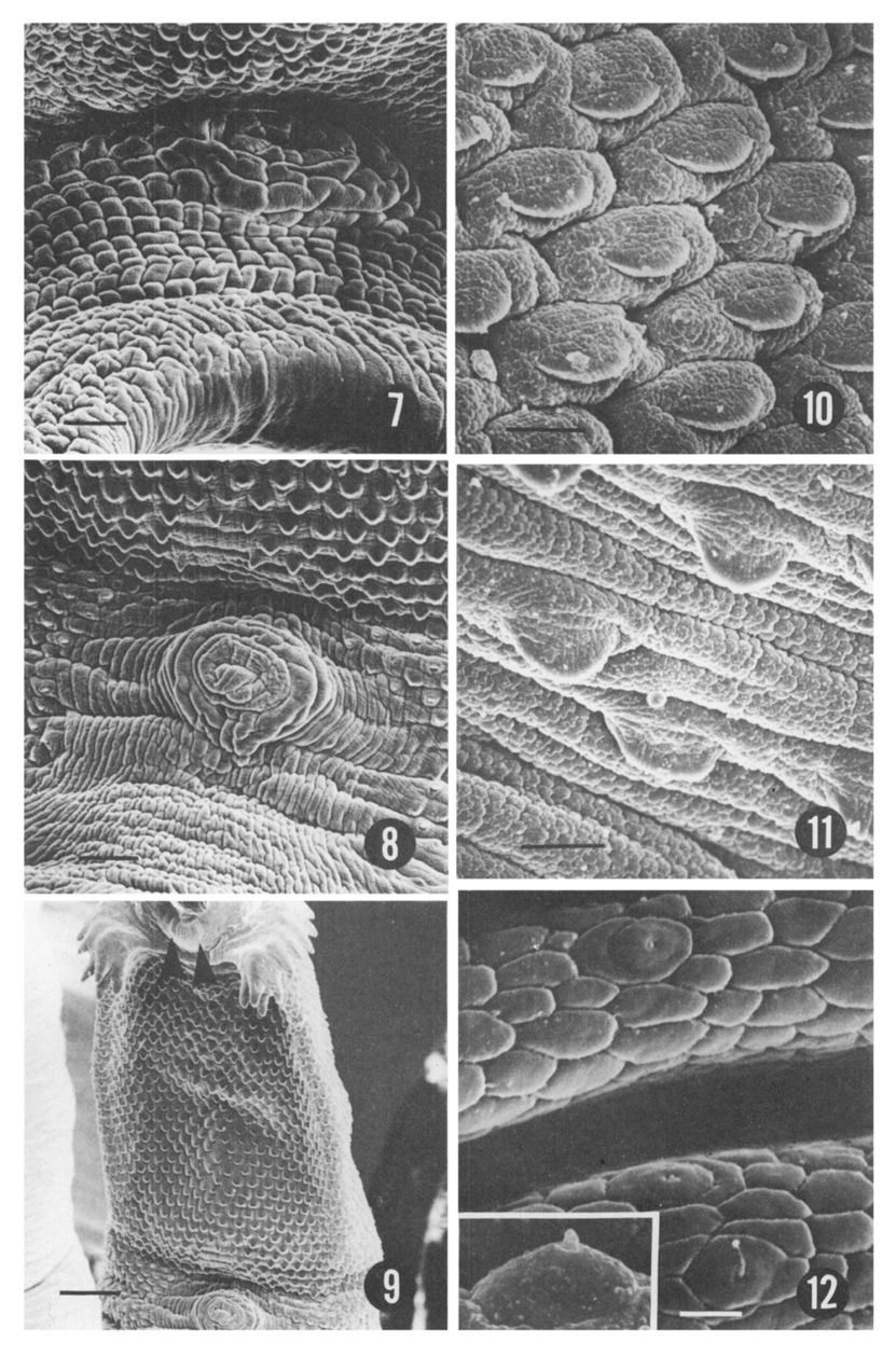
Figs. 7-12 Fig. 7. Tegumental surface around genital opening and ventral sucker where is free of trgumental spine. Bar=15µm.
Fig. 8. An erected cirrus protruding out from genital opening. Bar=20µm.
Fig. 9. Spines on the tegument between oral and ventral suckers which are arranged transversely and "X"-like pattern. Non-ciliated round swellings (type II papillae: arrow heads) on the lip of oral sucker are observed. Bar=50µm.
Fig. 10. Scale-like spines with round tip and broad base on the dorso-anterior tegument behind head crown. Bar=10µm.
Fig. 11. More sparsely distributed spines on the postero-ventral surface than in Fig. 10. Bar=10µm.
Fig. 12. Magnification of ciliated knob-like (type I) papillae on the tegument behind ventral sucker. Bar=2µm. Insert: A lateral view enlarged 2 times.
References
1.
Asada S. Trans Jap Pathol Soc 1926;16:293–294.
2.
Bennett CE. Scanning electron microscopy of Fasciola hepatica L. during growth and maturation in the mouse. J Parasitol 1975;61(5):892–898.
3.
Chai JY, Hong SJ, Sohn WM, Lee SH, Seo BS. Studies on intestinal tematodes in Korea: XVI. Infection status of loaches with the metacercariae of Echinostoma hortense. Korean J Parasitol 1985;23(1):18–23.
4.
Cho SY, Kang SY, Ryang YS. [Helminthes Infections In The Small Intestine Of Stray Dogs In Ejungbu City, Kyunggi Do, Korea]. Korean J Parasitol 1981;19(1):55–59.
5.
Fried B, Fujino T. Scanning electron microscopy of Echinostoma revolutum (Trematoda) during development in the chick embryo and the domestic chick. Int J Parasitol 1984;14(1):75–81.
6.
Fujino T, Ishii Y, Cho DW. Surface ultrastructure of the tegument of Clonorchis sinensis newly excysted juveniles and adult worms. J Parasitol 1979;65(4):579–590.
7.
Koie M. Stereoscan studies of cercariae, metacercariae, and adults of Cryptocotyle lingua (Creplin 1825) Fischoeder 1903 (Trematoda: Heterophyidae). J Parasitol 1977;63(5):835–839.
8.
Lee SH, Hong ST, Seo BS. [A Study On The Fine Tegumental Structures Of The Metacercaria And Juvenile Stages Of Clonorchis Sinensis]. Korean J Parasitol 1982;20(2):123–132.
9.
Lee SH, et al. Seoul J Med 1985;26(1):52–63.
10.
Lee SH, Seo BS, Chai JY, Hong SJ. [Study on Metagonimus yokogawai(Katsurada, 1912) in Korea VII. Electron microscopic observation on the tegumental structure]. Korean J Parasitol 1984;22(1):1–10.
11.
Lee SK, Chung NS, Ko IH, Ko HI, Chai JY. [Two cases of natural human infection by Echinostoma hortense]. Korean J Parasitol 1986;24(1):77–81.
12.
Lumsden RD. Surface ultrastructure and cytochemistry of parasitic helminths. Exp Parasitol 1975;37(2):267–339.
13.
Miyamoto K, et al. Japanese J Parasit 1983;32(4):261–269.
14.
Park JT. Keijo J Med 1938;9(4):283–286.
15.
Ryang YS, Ahn YK, Lee KW, Kim TS, Han MH. [Two cases of natural human infection by Echinostoma hortense and its second intermediate host in Wonju area]. Korean J Parasitol 1985;23(1):33–40.
16.
Seo BS, Cho SY, Hong ST, Hong SJ, Lee SH. Studies On Parasitic Helminths Of Korea 5.Survey On Intestinal Trematodes Of House Rats. Korean J Parasitol 1981;19(2):131–136.
17.
Seo BS, Hong ST, Chai JY, Lee SH. Studies On Intestinal Trematodes In Korea: VIII. A Human Case Of Echinostoma Hortense Infection. Korean J Parasitol 1983;21(2):219–223.
18.
Seo BS, Lee SH, Chai JY, Hong ST, Hong SJ. [Studies on intestinal trematodes in Korea X. Scanning electron microscopic observation on the tegument of Fibricola seoulensis]. Korean J Parasitol 1984;22(1):21–29.
19.
Seo BS, Rim HJ, Lee CW. Studies on the parasitic helmiths of Korea: I. Trematodes of rodents. Korean J Parasitol 1964;2(1):20–26.
20.
Smales LR, et al. J Helminth 1984;58:187–195.
21.
Tani S, et al. Japanese J Parasit 1974;23(6):404–408.
22.
Yoshida Y, et al. Japanese J Parasit 1986;35(2):62.