Warning: mkdir(): Permission denied in /home/virtual/lib/view_data.php on line 81
Warning: fopen(upload/ip_log/ip_log_2024-04.txt): failed to open stream: No such file or directory in /home/virtual/lib/view_data.php on line 83
Warning: fwrite() expects parameter 1 to be resource, boolean given in /home/virtual/lib/view_data.php on line 84 Purification of antigenic proteins of Paragonimus westermani and their applicability to experimental cat paragonimiasis
Purification of antigenic proteins of Paragonimus westermani and their applicability to experimental cat paragonimiasis
Won Young Choi,Jae Eul Yoo,Ho Woo Nam and Hyung Rak Choi
Catholic Institute of Parasitic Disease, Catholic Medical College, Korea.
Abstract
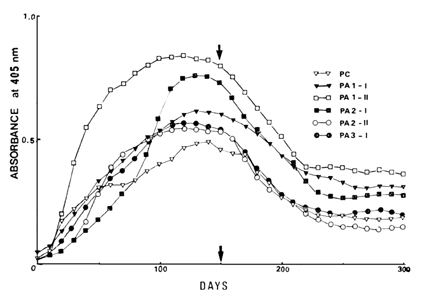
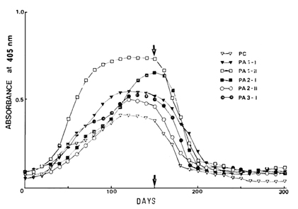
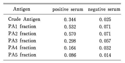
This study was designed to evaluate the partially purified antigens which were fractionated from crude extract of Paragonimus westermani and to monitor the enzyme-linked immunosorbent assay (ELISA) in experimental cat paragonimiasis during the course of infection as well as before and after chemotherapy. Crude extract of 6-month-old adult P. westermani was fractionated to 5 antigens by successive applications of ammonium sulfate precipitation, ion exchange chromatography and gel filtration. And the cats, 10 in each group, were infected with 60, 30, 15, and 5 metacercariae, then the half of each group was treated with praziquantel 2 times in one day of 100 mg per kilogram of weight on 150 days after the infection. Sera were collected every 10 days. ELISA was performed with the concentration of 2 µg/ml antigen, 100 times diluted sera and 1,000 times diluted alkaline phosphatase conjugated anti-cat IgG. The results were as follows: Absorbance by ELISA with proteins precipitated by differential concentration of ammonium sulfate was the highest at 51-65 per cent precipitate (PA2), followed by 0-50 per cent precipitate (PA1), 66-80 per cent precipitate (PA3), and 81-90 precipitate (PA4). Unprecipitated protein over 90 per cent ammonium sulfate (PA5) showed the lowest antigenicity. Fractionation of PA1, PA2, and PA3 through the DEAE-cellulose column did not differentiate the antigenic proteins. By passing through the Sephadex G-200 column, PAl and PA2 were fractionated to high molecular weight proteins and those of low molecular weight which showed high absorbance by ELISA (PA1-I, II and PA2-I, II). But PA3 was shown to have a fraction of high molecular weight proteins (PA3-I) which showed high antigenicity. SDS-polyacrylamide gel electrophoresis of PA1-I, PA1-II, PA2-I, PA2-II, PA3-I, and crude extract was performed. Fraction PA1-I was composed of proteins which had the molecular weight of 270 kilodaltons (KD) to 196 KD; of them 220 KD protein was major band. Fraction PA2-I was composed of 255-225 KD, and PA3-I, 255-240 KD, respectively. Fraction PA1-II and fraction PA2-II consisted of 30 KD proteins. Absorbance by ELISA began to increase within 10-20 days after the infection and reached the highest on 140-180 days, then made plateau thereafter. Absorbance by ELISA decreased after praziquantel treatment. In 60 metacercariae infection group, the absorbance had been decreasing, but remained within the positive range during observation period, while those of 30, 15, and 5 metacercariae infection groups turned to negative range. Fraction PA1-II showed the highest antigenicity in ELISA, then fraction PA2-I, fraction PA1-I , fraction PA2-II, fraction PA3-I and crude extract followed. In early phase of infection, the absorbance of fraction PA1-II showed more rapid increase than those of the other fractions and it came to positive range at 20-30 days after infection.
Figures
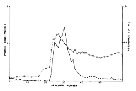
Fig. 1 Elution profile of PA1 (precipitated by 0~50% ammonium sulfate from crude antigen of P. Westermani) through DEAE-cellulose column and absorbance of ELISA for its fractions against a reference positive serum.
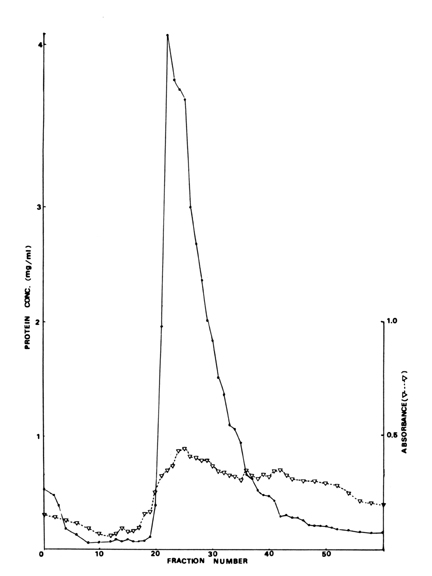
Fig. 2 Elution profile of PA2 (precipitated by 51~65% ammonium sulfate from crude antigen of P. Westermani) through DEAE-cellulose column and absorbance of ELISA for its fractions against a reference positive serum.
Fig. 3 Elution profile of PA3 (precipitated by 66~800% ammonium sulfate from crude antigen of P. Westermani) through DEAE-cellulose column and absorbance of ELISA for its fractions against a reference positive serum.
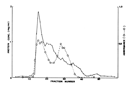
Fig. 4 Elution profile of PA1' through Sephadex G-200 gel and absorbance of ELISA from its fractions against a reference positive serum. PA1' is the fraction which is filtered PA1 through DEAE-cellulose column.
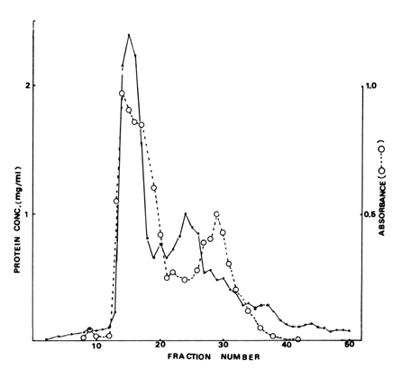
Fig. 5 Elution profile of PA2' through Sephadex G-200 gel and absorbance of ELISA from its fractions against a reference positive serum. PA2' is the fraction which is filtered PA2 through DEAE-cellulose column.
Fig. 6 Elution profile of PA3' through Sephadex G-200 gel and absorbance of ELISA from its fractions against a reference positive serum. PA3' is the fraction which is filtered PA3 through DEAE-cellulose column.
Fig. 7 SDS-PAGE patterns of crude and fractionated antigens.
C: crude antigen
D: fraction which is fractionated by DEAE-cellulose chromatography
I : fraction of high molecular weight proteins
II: fraction of low molecular weight proteins
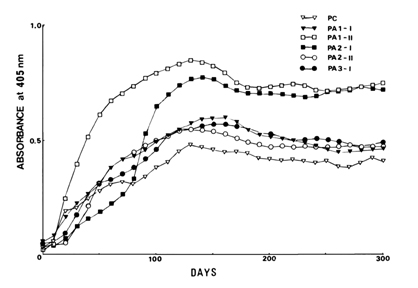
Fig. 8 Mean absorbance curves of ELISA against crude and fractionated antigens in the group of cats given 60 metacercariae.
Fig. 9 Mean absorbance curves of ELISA against crude and fractionated antigens in the group of cats given 30 metacercariae.
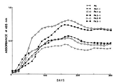
Fig. 10 Mean absorbance curves of ELISA against crude and fractionated antigens in the group of cats given 15 metacercariae.
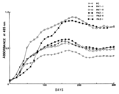
Fig. 11 Mean absorbance curves of ELISA against crude and fractionated antigens in the group of cats given 5 metacercariae.
Fig. 12 Mean absorbance curves of ELISA against crude and fractionated antigens in the group of cats given 60 metacercariae and treated with praziquantel.
Fig. 13 Mean absorbance curves of ELISA against crude and fractionated antigens in the group of cats given 30 metacercariae and treated with praziquantel.
Fig. 14 Mean absorbance curves of ELISA against crude and fractionated antigens in the group of cats given 15 metacercariae and treated with praziquantel.
Fig. 15 Mean absorbance curves of ELISA against crude and fractionated antigens in the group of cats given 5 metacercariae and treated with praziquantel.
Tables
Table 1 Absorbance values of ELISA for fractionated antigens by ammonium sulfate differential precipitation
References
1.
Ando A. Chugai Iji Simpo 1917;900:1122–1130.
2.
Ando A. Jpn J Microbiol Soc 1921;15:391–404.
3.
Capron A, et al. Ann Inst Past 1965;105:798–810.
4.
Chang HT, Wang CW, Yu CF, Hsu CF, Fang JC. Paragonimiasis; a clinical study of 200 adult cases. Chin Med J 1958;77(1):3–9.
5.
Cho SY, Hong ST, Rho YH, Choi SY, Han YC. Application of micro-ELISA in serodiagnosis of Human paragonimiasis. Korean J Parasitol 1981;19(2):151–156.
6.
Cho SY, Lee DK, Kang SY, Kim SI. [An Epidemiological Study Of Human Paragonimiasis By Means Of Micro-ELISA]. Korean J Parasitol 1983;21(2):246–256.
7.
Cho SY, Kim SI. [Detection Of Paragonimus-Specific Igg Antibody In Csf And Pleural Effusion By Micro-ELISA]. Korean J Parasitol 1983;21(2):286–288.
8.
Choi WY, Kimura K, Tsuji M. [Immunoelectrophoresis For Anthelmintics Evaluation Against Experimental Paragonimiasis]. Korean J Parasitol 1976;14(2):94–102.
9.
Engvall E, Perlmann P. Enzyme-linked immunosorbent assay (ELISA). Quantitative assay of immunoglobulin G. Immunochemistry 1971;8(9):871–874.
10.
Grabar P, Williams CA. [Method permitting the combined study of the electrophoretic and the immunochemical properties of protein mixtures; application to blood serum]. Biochim Biophys Acta 1953;10(1):193–194.
11.
Kim JW, et al. J Med Center 1964;5:3–4.
12.
Kim CH, et al. Chung-Ang J med 1982;7:335–347.
13.
Kim SI, Kang SY, Cho SY. [On The Applicability Of Partially Purified Antigenic Preparations Of Paragonimus Westermani]. Korean J Parasitol 1983;21(2):257–264.
14.
Lee OR, Choi WY. [Comparison Of Agar-Gel Diffusion Tests, Counterimmunoelectrophoresis And Enzyme-Linked Immunosorbent Assay In The Sera Of Skin Test Positives For Paragonimiasis]. Korean J Parasitol 1983;21(2):270–280.
15.
Lee YK, Ryu JS, Lee KT, Im KI. [Comparison Of Tia With ELISA For Circulating Antibody Detection In Clonorchiasis]. Korean J Parasitol 1983;21(2):265–269.
16.
Lowry OH, Rosebrough NJ, Farr AL, Randall RJ. Protein measurement with the Folin phenol reagent. J Biol Chem 1951;193(1):265–275.
17.
McLaren M, Draper CC, Roberts JM, Minter-Goedbloed E, Ligthart GS, Teesdale CH, Amin MA, Omer AH, Bartlett A, Voller A. Studies on the enzyme linked immunosorbent assay (ELISA) test for Schistosoma mansoni infections. Ann Trop Med Parasitol 1978;72(3):243–253.
18.
Merril CR, Goldman D, Sedman SA, Ebert MH. Ultrasensitive stain for proteins in polyacrylamide gels shows regional variation in cerebrospinal fluid proteins. Science 1981;211(4489):1437–1438.
19.
Min DY, Soh CT. Evaluation Of Specific Ige Antibody In Clonorchis Sinensis Infection. Korean J Parasitol 1983;21(1):27–31.
20.
Ouchterlony O. Diffusion-in-gel methods for immunological analysis. Prog Allergy 1958;5:1–78.
21.
Sawada T, Takei K, Voneyama K. Studies On The Immunodiagnosis Of Paragonimiasis I The Precipitin Reaction With Crude And Fractionated Antigens. J Infect Dis 1964;114:311–314.
22.
Speiser F. Application of the enzyme-linked immunosorbent assay (ELISA) for the diagnosis of filariasis and echinococcosis. Tropenmed Parasitol 1980;31(4):459–466.
23.
Yogore MG, et al. Am J Trop Med Hyg 1965;14:586–591.
24.
Yogore MG Jr, Lewert RM, Blas BL. Schistosomiasis japonica in Barrio San Antonio, Basey, Samar, in the Philippines V The enzyme-linked immunosorbent assay (ELISA) compared with quantitative stool examination and the circumoval precipitin (COP) test. Am J Trop Med Hyg 1981;30(6):1252–1262.