Warning: mkdir(): Permission denied in /home/virtual/lib/view_data.php on line 81
Warning: fopen(upload/ip_log/ip_log_2024-04.txt): failed to open stream: No such file or directory in /home/virtual/lib/view_data.php on line 83
Warning: fwrite() expects parameter 1 to be resource, boolean given in /home/virtual/lib/view_data.php on line 84 Specific IgG antibody responses in experimental cat metagonimiasis
Specific IgG antibody responses in experimental cat metagonimiasis
Seung-Yull Cho,Suk Il Kim and Shin Yong Kang
Department of Parasitology, College fo Medicine, Chung-Ang University, Seoul 151, Korea.
Abstract
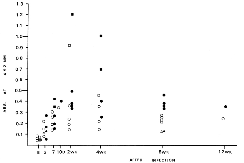
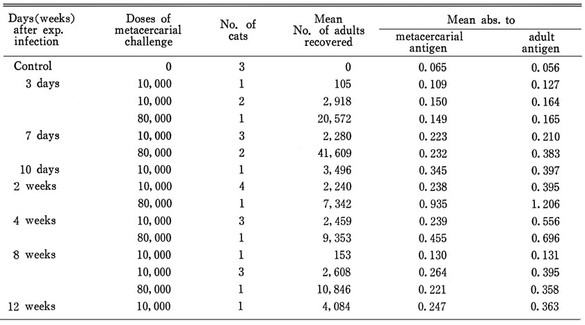
In order to observe the feasibility of serologic diagnosis of metagonimiasis, saline extracts of metacercariae and 4-week old adults were prepared. Sera from 25 experimentally infected cats were collected from 3 days to 12 weeks after infection. Their levels of specific IgG antibody were measured by ELISA together with 3 sera from non-infected cats. Specific IgG antibody levels began to rise in 7 days after infection, reached their peak in 2-4 weeks and made a plateau thereafter. Cats infected with hundreds of adult worms showed minimal rise of the antibody level. Adult antigen was superior to metacercarial antigen in detecting the specific IgG antibody.
Figures
Fig. 1 Specific IgG antibody levels in individual cat as measured by ELISA. Open triangle, circle and rectangle (▵, ◦, ▫) mean antibody levels to metacercarial antigen while closed ones (▴, •, ▪) mean those to adult antigen. Triangles (▵,▴): cats infected with less than 1,000, circles (◦, •): cats infected with 1,000~5,000, rectangles (▫, ▪): cats infected with more than 5,000 adults. Markings (◦, ▫) of B (in control cats) represents abs. to metacercarial (◦) or adult (▫) antigens.
Tables
Table 1 Specific IgG antibody levels as measured by ELISA in 25 experimental cats infected with different doses of Metagonimus yokogawai
References
1.
Chai JY. Seoul J Med 1979;20(2):104–117.
2.
Han JH, et al. Korea Univ Med J 1986;23:13–25.
3.
Kang SY, Cho SY, Chai JY, Lee JB, Jang DH. A Study On Intestinal Lesions Of Experimentally Reinfected Dogs With Metagonimus Yokogawai. Korean J Parasitol 1983;21(1):58–73.
4.
Kim ER, et al. Chung-Ang J Med 1985;10(3):291–306.
5.
Lee JB, Chi JG, Lee SK, Cho SY. Study On The Pathology Of Metagonimiasis In Experimentally Infected Cat Intestine. Korean J Parasitol 1981;19(2):109–129.
6.
Lowry OH, Rosebrough NJ, Farr AL, Randall RJ. Protein measurement with the Folin phenol reagent. J Biol Chem 1951;193(1):265–275.
7.
McLaren M, Draper CC, Roberts JM, Minter-Goedbloed E, Ligthart GS, Teesdale CH, Amin MA, Omer AH, Bartlett A, Voller A. Studies on the enzyme linked immunosorbent assay (ELISA) test for Schistosoma mansoni infections. Ann Trop Med Parasitol 1978;72(3):243–253.