Warning: mkdir(): Permission denied in /home/virtual/lib/view_data.php on line 81
Warning: fopen(upload/ip_log/ip_log_2024-04.txt): failed to open stream: No such file or directory in /home/virtual/lib/view_data.php on line 83
Warning: fwrite() expects parameter 1 to be resource, boolean given in /home/virtual/lib/view_data.php on line 84 Experimental life history of Echinostoma hortense
S H Lee,S W Hwang,W M Sohn,*W G Kho,*S T Hong and J Y Chai
Department of Parasitology and Institute of Endemic Diseases, Seoul National University College of Medicine, Seoul 110-460, Korea.
Abstract
The complete life cycle of Echinostoma hortense has been maintained in the laboratory, using Lymnaea pervia snails and Rana nigromaculata tadpoles as the first and second intermediate hosts. ICR mice was used as the definitive host. Within the egg of E. hortense, the miracidium was fully matured in 13 days of incubation at 29-30℃. The miracidium was 93.8 × 53.6 µm in average size, covered with numerous cilia of 7-11 µm length. The epidermal plates were arranged in 6-8-4-2 formula. The first generation rediae (1.19 × 0.27 mm in average size) were observed in 14 days after miracidial challenge to the snails, and the second generation rediae (1.40 × 0.26 mm in average size) in 30 days. The average size of the cercaria was 295.5 × 145.0 µm. Their head crown was poorly developed, and collar spines were not yet observed.
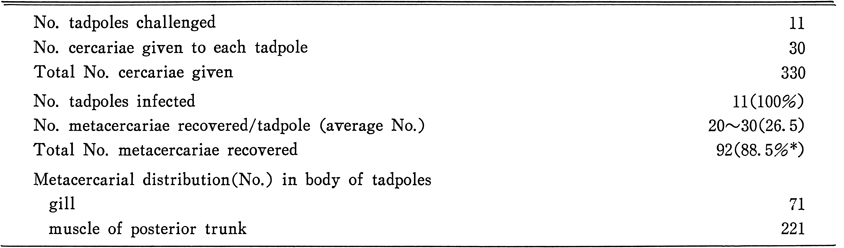
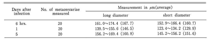
After a cercarial challenge to the tadpoles, all of the tadpoles became infected and the average worm recovery rate was 88.5%. The majority of the metacercariae (75.5%) were recovered from the muscle of the tadpole's posterior body and the rest (24.3%) from their gills. The metacercariae from the tadpoles were elliptical, and 167.7 × 129.9 µm in average size. The recovery rate of adults from the mice was different by the age of the metacercariae grown in the tadpoles. The metacercariae younger than 5 hrs could not infect mice whereas those older than 6 hrs could infect mice. The recovery rate became higher as the metacercaria matured, with the peak recovery rate of 90.0% at the metacercarial age of 9 days. Thereafter the recovery rate decreased to 55.0% at the age of 50 days. As shown by the above results, the whole life cycle of E. hortense has been completed in the laboratory. At least 55-58 days were required to maintain one egg-to-egg cycle of E. hortense.
Figures
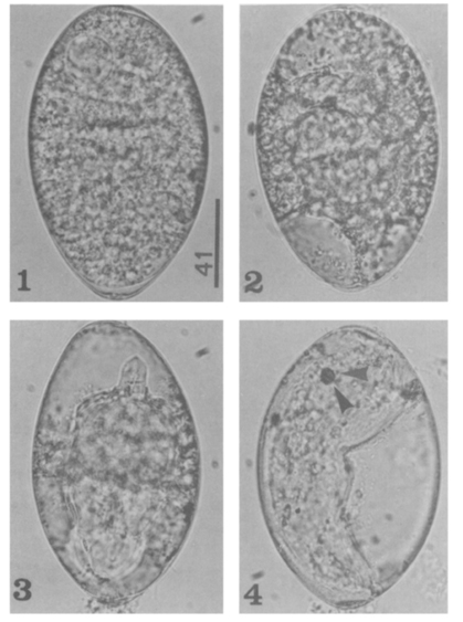
Figs. 1-4 Fig. 1. A newly laid egg of Echinostoma hortense.
Fig. 2. A developing egg after 6 day of incubation.
Fig. 3. A developing egg after 8 day of incubation.
Fig. 4. An almost fully-developed egg after 13 days of incubation.
*Bar unit: µm
Fig. 5 A miracidium of E. hortense showing an eye spot and numerous cilia.
*Bar unit: µm
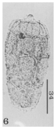
Fig. 6 A miracidium of E. hortense showing epidermal plates.
*Bar unit: µm
Fig. 7 Lymnaea pervia, used as the first intermediate host in this study.
*Bar unit: µm
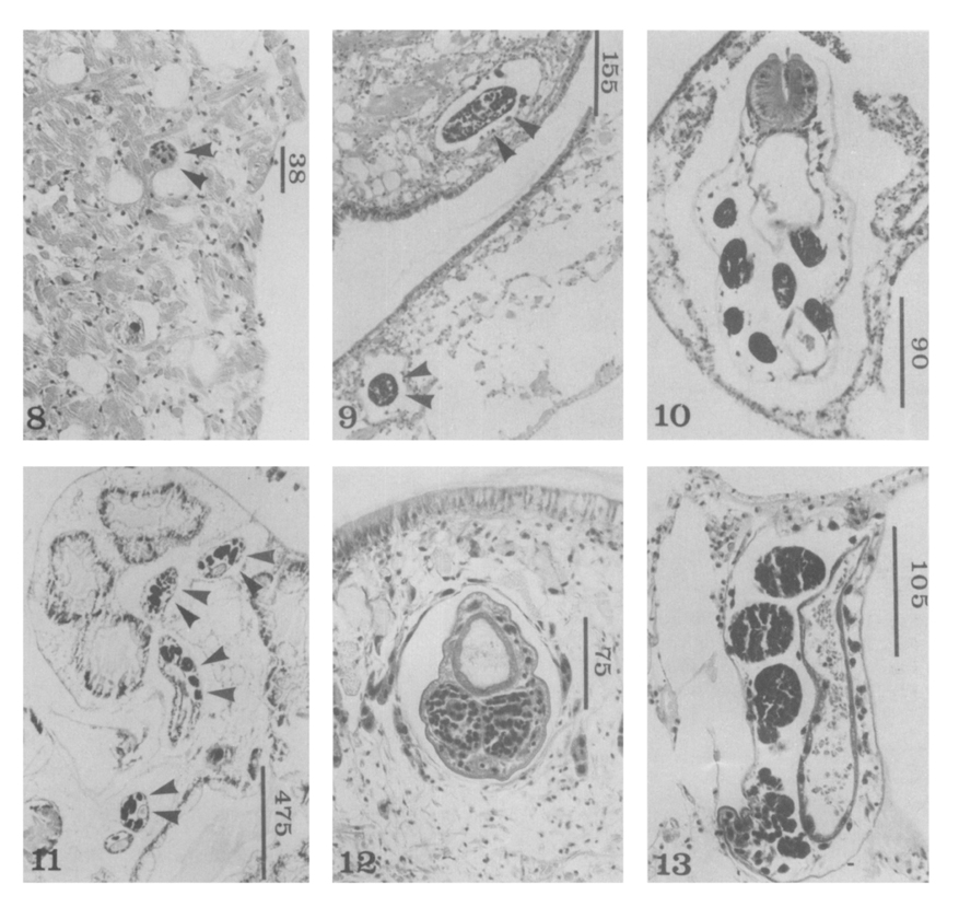
Figs. 8-13 Fig. 8. Sporocysts of E. hortense (arrow heads) in the mantle of a snail 2 hrs after miracidial infection.
Fig. 9. Sporocysts of E. hortense (arrow heads) in the head-foot of a snail 7 days after miracidial challenge.
Fig. 10. A redia in the cephalopedal sinus of a snail, 16 days old.
Fig. 11. Rediae (arrow heads) in the digestive glands of a snail, 16 days old.
Fig. 12. A redia in the head-foot of a snail, 21 days old.
Fig. 13. A redia in the hemocele of a snail, 21 days old.
*Bar unit: µm
Figs. 14-17 Fig. 14. A first generation redia, 15 day old.
Fig. 15. A Second generation redia, 25 day old.
Fig. 16. A Second generation redia, 30 day old.
Fig. 17. A cercaria obtained from an experimental snail.
*Bar Unit: µm
Figs. 18-22 Fig. 18. Metacercariae in the gill of a tadpole.
Fig. 19. A metacercaria isolated from the gill of a tadpole.
Fig. 20. An excysted metacercaria showing the collar spines and excretory granules.
Fig. 21. A three-day old juvenile worm showing 4 end group spines (arrow heads).
Fig. 22. Adult of E. hortense from an experimental nouse 12 days after infection.
*Bar unit: µm
Tables
Table 1 Measurements of E. hortense rediae from experimentally infected snails
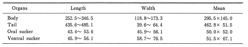
Table 2 Measurements* of E. hortense cercariae from experimentally infected snails (on micrometers)
Table 3 Recovery rates of E. hortense metacercariae from experimentally infected tadpoles
Table 4 Measurements of E. hortense metacercariae from experimentally infected tadpoles
Table 5 The infectivity of E. hortense metacercariae by the age of infection in tadpoles
Table 6 The period form development of E. hortense by stage
References
1.
Ahn YK, Ryang YS. [Experimental and epidemiological studies on the life cycle of Echinostoma hortense Asada, 1926 (Trematoda: Echinostomatidae)]. Korean J Parasitol 1986;24(2):121–136.
2.
Ahn YK, Ryang YS, Chung PR, Lee KT. [Echinostoma hortense metacercariae naturally encysted in Odontobutis obscura interrupta (a freshwater fish) and experimental infection to rats]. Korean J Parasitol 1985;23(2):230–235.
3.
Alicata JE. Life cycle and developmental stages of Philophthalmus gralli in the intermediate and final hosts. J Parasitol 1962;48:47–54.
4.
Arizono N, et al. Jpn J Parasitol 1976;25(1):36–45.
5.
Asada S. Trans Jap Pathol Soc 1926;16:293–294.
6.
Asada S. Vol Jub Yoshida 1939;1:39–69.
7.
Chai JY, Hong SJ, Sohn WM, Lee SH, Seo BS. Studies on intestinal tematodes in Korea: XVI. Infection status of loaches with the metacercariae of Echinostoma hortense. Korean J Parasitol 1985;23(1):18–23.
8.
Cho SY, Kang SY, Ryang YS. [Helminthes Infections In The Small Intestine Of Stray Dogs In Ejungbu City, Kyunggi Do, Korea]. Korean J Parasitol 1981;19(1):55–59.
9.
Eom KS, Rim HJ, Jang DH. A Study On The Parasitic Helminths Of Domestic Duck(Anas Platyrhynchos Var. Domestica Linnaeus) In Korea. Korean J Parasitol 1984;22(2):215–221.
10.
Fashuyi SA. Acta Parasitol Polon 1985;30:1–9.
11.
Hirasawa I. Kumamoto Igakkai Zashi 1929;5:599.
12.
Chu JK, Cho YJ, Chung SB, Won BO, Yoon MB. [Study on the trematode parasites of the birds in Korea]. Korean J Parasitol 1973;11(2):70–75.
13.
Itagaki T, et al. Jpn J Parasitol 1986;35(6):505–511.
14.
Kawanaka M. Jpn J Parasitol 1978;27:214–224.
15.
Lee SH, Sohn WM, Chai JY. Echinostoma revolutum and Echinoparyphium recurvatum recovered from house rats in Yangyang-gun, Kangwon-do. Korean J Parasitol 1990;28(4):235–240.
16.
Lee SH, Shin SM, Hong ST, Sohn WM, Chai JY, Seo BS. Growth and development of Fibricola seoulensis metacercariae in tadpoles. Korean J Parasitol 1986;24(2):109–114.
17.
Lee SK, Chung NS, Ko IH, Ko HI, Chai JY. [Two cases of natural human infection by Echinostoma hortense]. Korean J Parasitol 1986;24(1):77–81.
18.
Lee SK, Chung NS, Ko IH, Sohn WM, Hong ST, Chai JY, Lee SH. [An epidemiological suryey of Echinostoma hortense infection in Chongsong-gun, Kyongbuk province]. Korean J Parasitol 1988;26(3):199–206.
19.
Mori J. Tokyo Iji Shinshi 1935;2929:1237–1244.
20.
Okahashi K. Okayama Igakkai Zasshi 1966;78:15–24.
21.
Ono S. Dobutsugaku Zasshi 1930;42:7–16.
22.
Pan CT. Studies on the host-parasite relationship between Schistosoma mansoni and the snail Australorbis glabratus. Am J Trop Med Hyg 1965;14(6):931–976.
23.
Park JT. Keijo J Med 1938;9(4):283–286.
24.
Ryang YS, Ahn YK, Lee KW, Kim TS, Han MH. [Two cases of natural human infection by Echinostoma hortense and its second intermediate host in Wonju area]. Korean J Parasitol 1985;23(1):33–40.
25.
Ryang YS, Ahn KY, Kim WT, Shin KC, Lee KW, Kim TS. [Two cases of human infection by Echinostoma cinetorchis]. Korean J Parasitol 1986;24(1):71–76.
26.
Saito S, et al. Jpn J Parasitol 1982;31(4):281–287.
27.
Seo BS, Lee SH, Chai JY, Hong SJ. Studies on intestinal trematodes in Korea XX. Four cases of natural human infection by Echinochasmus japonicus. Korean J Parasitol 1985;23(2):214–220.
28.
Seo BS, et al. Seoul J Med 1980;21(1):21–29.
29.
Seo BS, Cho SY, Hong ST, Hong SJ, Lee SH. Studies On Parasitic Helminths Of Korea 5.Survey On Intestinal Trematodes Of House Rats. Korean J Parasitol 1981;19(2):131–136.
30.
Seo BS, Hong ST, Chai JY, Lee SH. Studies On Intestinal Trematodes In Korea: VIII. A Human Case Of Echinostoma Hortense Infection. Korean J Parasitol 1983;21(2):219–223.
31.
Seo BS, Rim HJ, Lee CW. Studies on the parasitic helmiths of Korea: I. Trematodes of rodents. Korean J Parasitol 1964;2(1):20–26.