Warning: mkdir(): Permission denied in /home/virtual/lib/view_data.php on line 81
Warning: fopen(upload/ip_log/ip_log_2024-04.txt): failed to open stream: No such file or directory in /home/virtual/lib/view_data.php on line 83
Warning: fwrite() expects parameter 1 to be resource, boolean given in /home/virtual/lib/view_data.php on line 84 Correlation of sonographic findings with histopathological changes of the bile ducts in rabbits infected with Clonorchis sinensis
Correlation of sonographic findings with histopathological changes of the bile ducts in rabbits infected with Clonorchis sinensis
S T Hong,1K H Park,2M Seo,1B I Choi,3J Y Chai,1 and S H Lee*1
1Department of Parasitology and Institute of Endemic Diseases, Seoul National University College of Medicine, Seoul 110-799, Korea.
Received October 10, 1994; Accepted November 21, 1994.
Abstract
Clonorchiasis is an important parasitic disease of humans in Korea. The present study intended to compare sonographic findings with histopathological changes in experimental clonorchiasis. Eighteen New Zealand white rabbits were infected with metacercariae of Clonorchis sinensis, and examined 4, 10, and 22 weeks post-infection (PI). Four infected rabbits were treated with praziquantel 10 weeks PI and were examined 12 weeks after treatment. Sonography revealed mild to severe dilatation of the intrahepatic ducts (IHDD) and slightly increased periductal echoes in 12 out of 14 rabbits at 4 weeks PI, and all of the animals after 10 and 22 weeks PI and 12 weeks after treatment. The histopathological lesions were duct dilatation, mucosal hyperplasia, and periductal fibrosis, which progressed from 4 weeks to 22 weeks PI and even in treated rabbits. The dilated intrahepatic ducts over 1 mm diameter were detected by sonography. The present results indicate that sonographic findings are well correlated with histopathological lesions in rabbit clonorchiasis except for early phase of light burden of infection. The sonography has a limitation in discriminating residual sequelae of the ducts after praziquantel treatment.
Figures
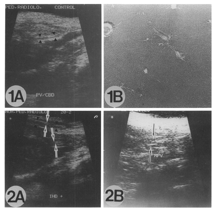
Figs. 1-2 Fig. 1A. Sonogram of a control rabbit liver. The portal vein branches (arrow heads) are visualized but bile ducts are not. Fig. 1B. Histopathological observation of the liver of the control rabbit, showing normal architecture of the liver and normal intrahepatic ducts. HE stain, original magnification × 60. Fig. 2A. sonogram of the rabbit 20-2 at 4 weeks PI. The portal vein (arrow heads) and bile ducts (arrows) were visualized. Fig. 2B. Sonogram of the rabbit 20-2. 10 weeks PI. showing moderate dilatation of the intrahepatic ducts (arrows) along the portal vein and increased periductal echoes.
Figs. 3-4 Fig. 3A. Sonogram of the 20-4 rabbit, 10 weeks PI. The ducts (arrows) are dilated moderately with periductal echoes. Fig. 3B. Photomicrograph of the rabbit 20-4, 10 weeks PI. The ducts (arrows) infected by flukes are remarkably dilated. Portal vein (arrow heads) is also seen nearby. HE stain, original magnification × 15. Fig. 4A. Sonogram of the 400-15 rabbit, 12 weeks after treatment. The ducts (arrows) are still dilated moderately with increased periductal echoes. Fig. 4B. Photomicrograph of the rabbit 400-15, 12 weeks after treatment. The ducts (arrows) are still dilated. Portal vein (arrow heads) is also seen nearby. HE stain, original magnification × 15.
Tables
Table 1 Scheme of experimental infection of rabbits with Clonorchis sinensis
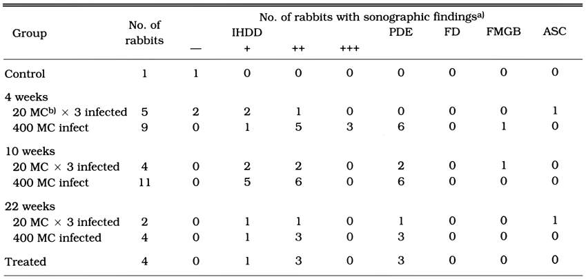
Table 2 Sonographic findings of rabbits 4, 10 and 22 weeks after infection with C. sinensis
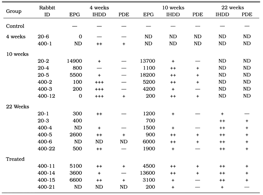
Table 3 EPG and sonographic findings of rabbits 4, 10 and 22 weeks after infection with C. sinensis
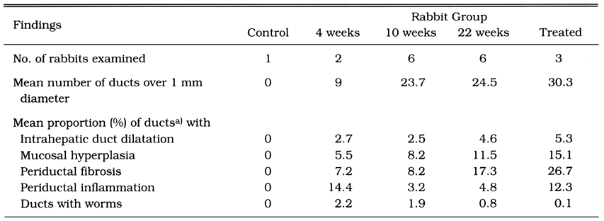
Table 4 Histopathological findings of rabbits infected with C. sinensis
References
1.
Chen M, Lu Y, Hua X, Mott KE. Trop Dis Bull 1994;91:R7–R65.
2.
Choi BI, Kim HJ, Han MC, Do YS, Han MH, Lee SH. CT findings of clonorchiasis. Am J Roentgenol 1989;152:281–284.
3.
Choi BI, Kim HJ, Han MC, Do YS, Han MH, Lee SH. CT findings of clonorchiasis. AJR Am J Roentgenol 1989;152(2):281–284.
4.
Hong ST, Kho WG, Kim WH, Chai JY, Lee SH. Turnover of biliary epithelial cells in Clonorchis sinensis infected rats. Korean J Parasitol 1993;31(2):83–89.
5.
Kang IW, Seo HS, Lim DR, Yeon KM. J Korean Radiol Soc 1980;16(1):159–162.
6.
Kim JW, Kim JG, Sol CH, Kim BS. J Korean Radiol Soc 1983;19(3):538–545.
7.
Kim MJ, Yoo HS, Lee JT, Jung SH. J Korean Radiol Soc 1988;24(5):878–882.
8.
Lee JH, Rim HJ, Bak UB. Effect of Clonorchis sinensis infection and dimethylnitrosamine administration on the induction of cholangiocarcinoma in Syrian golden hamsters. Korean J Parasitol 1993;31(1):21–30.
9.
Lee SH, Hong ST, Kim CS, Sohn WM, Chai JY, Lee YS. Histopathological changes of the liver after praziquantel treatment in Clonorchis sinensis infected rabbits. Korean J Parasitol 1987;25(2):110–122.
10.
Lee SH, Chai JY, Yang EC, Yun CK, Hong ST, Lee JB. Seoul J Med 1988;29(3):253–262.
11.
Lee SH, Lee JI, Huh S, Yu JR, Chung SW, Chai JY, Hong ST. Secretions of the biliary mucosa in experimental clonorchiasis. Korean J Parasitol 1993;31(1):13–20.
12.
Lee SH, Shim TS, Lee SM, Chi JG. [Studies On Pathological Changes Of The Liver In Abino Rats Infected With Clonorchis Sinensis]. Korean J Parasitol 1978;16(2):148–155.
13.
Lim JH. Radiologic findings of clonorchiasis. AJR Am J Roentgenol 1990;155(5):1001–1008.
14.
Lim JH, Ko YT, Kim SY, Ryu HS. J Korean Radiol Soc 1984;20:644–647.
15.
Lim JH, Ko YT, Lee DH, Min YI. J Korean Soc Med Ultrasound 1987;6(2):193–194.
16.
Lim JH, Ko YT, Lee DH, Kim SY. Clonorchiasis: sonographic findings in 59 proved cases. AJR Am J Roentgenol 1989;152(4):761–764.
17.
Morikawa P, Ishida H, Niizawa M, Komatsu M, Arakawa H, Masamune O. Sonographic features of biliary clonorchiasis. J Clin Ultrasound 1988;16(9):655–658.
18.
Rim HJ. The current pathobiology and chemotherapy of clonorchiasis. Korean J Parasitol 1986;24 Suppl:1–141.
19.
Ryu KN, Lim JH, Cho YJ, Yang MH. J Korean Radiol Soc 1993;29(1):1–8.
20.
Seo BS, Lee SH, Cho SY, Chai JY, Hong ST, Han IS, Sohn JS, Cho BH, Ahn SR, Lee SK, Chung SC, Kang KS, Shim HS, Hwang IS. An Epidemiologic Study On Clonorchiasis And Metagonimiasis In Riverside Areas In Korea. Korean J Parasitol 1981;19(2):137–150.
21.
Song GA, Kim JD, Lee DW, et al. Korean J Int Med 1989;37(3):344–355.