Warning: mkdir(): Permission denied in /home/virtual/lib/view_data.php on line 81
Warning: fopen(upload/ip_log/ip_log_2024-04.txt): failed to open stream: No such file or directory in /home/virtual/lib/view_data.php on line 83
Warning: fwrite() expects parameter 1 to be resource, boolean given in /home/virtual/lib/view_data.php on line 84 The effect of silica on the development of experimental Acanthamoeba meningoencephalitis with reference to the macrophage role in mice
The effect of silica on the development of experimental Acanthamoeba meningoencephalitis with reference to the macrophage role in mice
H S Lee,H J Shin,M S La and K Im*
Department of Parasitology, Yonsei University College of Medicine and Institute of Tropical Medicine, Yonsei University , Seoul 120-752, Korea.
Received August 24, 1994; Accepted October 11, 1994.
Abstract
The role of macrophages was observed in intranasally infected C3H/HeJ mice with trophozoites (3 × 10⁵) of Acanthamoeba culbertsoni which was a kind of free-living amoebae inducing meningoencephalitis in human and experimental animals. The mortality was 60% in the group of intraperitoneally injected mice with silica (0.5 mg/0.5 ml). It was much higher than that of 10% in the group of amoeba infected mice without silica administration. The phagocytic index of peritoneal macrophages co-cultured with Toxoplasma gondii was estimated daily. In contrast to the control and amoeba infected group which didn't show significant fluctuation of the phagocytic indices, the silica administrated group revealed under 3% until day 3, and gradual increase up to 24.7% in day 5 which was same level of amoeba infected group without silica administration. The level of interleukin-1b (IL-1b) measured by ELISA was the highest in the amoeba infected group without silica injection and the lowest in the amoeba infected group with silica administration. In the test of the amoebicidal activity of mice peritoneal macrophages in vitro, silica administration revealed reducing effect on amoebicidal activity of macrophages. In conclusion, macrophages were proven to play a significant role in defense mechanism against the development of experimentally induced Acanthamoeba meningoencephalitis.
Figures
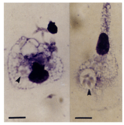
Fig. 1 Toxoplasma gondii tachyzoites (arrowhead) are observed in the cytoplasm of mouse peritoneal macrophages (Scale: 10 µm, Giemsa stain).
Fig. 2 Mouse peritoneal macrophages and Acanthamoeba culbertsoni trophozoites were cocultured for 2 hours in RPMI 1640 medium (Scale: 10 µm, Giemsa stain). A: A, culbertsoni trophozoites M: peritoneal macrophages.
Tables
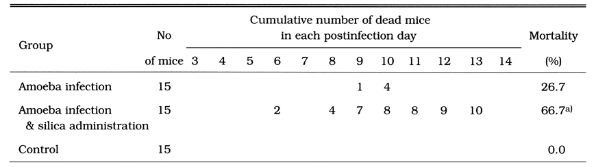
Table 1 Mortality of Aconthamoeba culbertsoni-infected mice, which were injected with silica 0.5 mg intraperitoneally on day 3 before infection
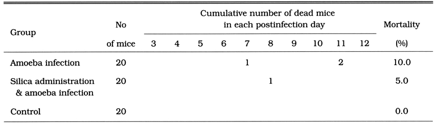
Table 2 Mortality of mice infected with Acanthamoeba culbertsoni and injected with silica 0.5 mg at the same time
Table 3 Mortality of mice infected with Acanthamoeba culbertsoni and injected with silica 0.25 mg at the same time
Table 4 Phagocytic index of mouse peritoneal macrophages on each observation day after Acanthamoeba culbertsoni infection
Table 5 Amoebicidal activity of mouse peritoneal macrophages against Acanthamoeba culbertsoni trophozoites. These two kinds of cells were cocultured for 2 days
Table 6 Interleukin-lβ level in serum of mice infected with Acanthamoeba culbertsoni and administered with slica
References
1.
Cleary SF, Marciano-Cabral F. Activated macrophages demonstrate direct cytotoxicity, antibody-dependent cellular cytotoxicity, and enhanced binding of Naegleria fowleri amoebae. Cell Immunol 1986;98(1):125–136.
2.
Cursons RT, Brown TJ, Keys EA, Moriarty KM, Till D. Immunity to pathogenic free-living amoebae: role of cell-mediated immunity. Infect Immun 1980;29(2):408–410.
3.
Ferrante A, Rowan-Kelly B. Activation of the alternative pathway of complement by Acanthamoeba culbertsoni. Clin Exp Immunol 1983;54(2):477–485.
4.
Fischer-Stenger K, Cabral GA, Marciano-Cabral F. Separation of soluble amoebicidal and tumoricidal activity of activated macrophages. J Protozool 1992;39(1):235–241.
5.
Fischer-Stenger K, Cabral GA, Marciano-Cabral F. The interaction of Naegleria fowleri amoebae with murine macrophage cell lines. J Protozool 1990;37(3):168–173.
6.
Gery I, Davies P, Derr J, Krett N, Barranger JA. Relationship between production and release of lymphocyte-activating factor (interleukin 1) by murine macrophages. 1. Effects of various agents. Cell Immunol 1981;64(2):293–303.
7.
Hilgers LA, Snippe H, Jansze M, Willers JM. Effect of in vivo administration of different adjuvants on the in vitro candidacidal activity of mouse peritoneal cells. Cell Immunol 1985;90(1):14–23.
8.
Kumazawa Y, Takimoto H, Nishimura C, Kawakita T, Nomoto K. Activation of murine peritoneal macrophages by saikosaponin a, saikosaponin d and saikogenin d. Int J Immunopharmacol 1989;11(1):21–28.
9.
Lallinger GJ, et al. Immunology 1987;55:1289–1293.
10.
Le J, Vilcek J. Tumor necrosis factor and interleukin 1: cytokines with multiple overlapping biological activities. Lab Invest 1987;56(3):234–248.
11.
Lefkowitz DL, et al. Methods in Enzymology 1986;32:537–542.
12.
Makioka A, et al. Jpn J Parasitol 1983;32(3):203–210.
13.
Martinez AJ. Infection of the central nervous system due to Acanthamoeba. Rev Infect Dis 1991;13 Suppl 5:S399–S402.
14.
Newton RC, Dowling R, Daulerio AJ, Culp S. An ELISA assay for murine interleukin-1 beta. J Immunol Methods 1993;161(2):257–264.
15.
Papaccio G, Frascatore S, Esposito V, Pisanti FA. Early macrophage infiltration in mice treated with low-dose streptozocin decreases islet superoxide dismutase levels: prevention by silica pretreatment. Acta Anat (Basel) 1991;142(2):141–146.
16.
Park YH, Osmond DG. Regulation of early precursor B cell proliferation in mouse bone marrow: stimulation by exogenous agents mediated by macrophages in the spleen. Cell Immunol 1991;135(1):168–183.
17.
Ringsted J, Jager BV, Suk D, Visvesvara GS. Probable acanthamoeba meningoencephalitis in a Korean child. Am J Clin Pathol 1976;66(4):723–730.
18.
Stern JJ, Graybill JR, Drutz DJ. Murine amebiasis: the role of the macrophage in host defense. Am J Trop Med Hyg 1984;33(3):372–380.
19.
Thong YH, Ferrante A, Shepherd C, Rowan-Kelly B. Resistance of mice to Naegleria meningoencephalitis transferred by immune serum. Trans R Soc Trop Med Hyg 1978;72(6):650–652.
20.
Voller A, Bidwell DE, Bartlett A. Enzyme immunoassays in diagnostic medicine. Theory and practice. Bull World Health Organ 1976;53(1):55–65.
21.
Zidek Z, Frankova D, Masek K. Some cellular and pathophysiological correlates of the inflammatory effects of a synthetic immunomodulatory agent, muramyl dipeptide (MDP). Agents Actions 1993;38(1-2):106–115.