Malaria control still remains a challenge in Africa where 45 countries, including Nigeria, are endemic for malaria, and about 588 million people are at risk [1]. The protection of pregnant women living in malaria-endemic countries has been of particular interest to many National Malaria Control Programmes because of their reduced immunity. Most cases of malaria in pregnancy in areas of stable malaria transmission are asymptomatic [2,3]. This is attributed to anti-disease immunity acquired during previous exposures which protects against clinical malaria [4]. Unfortunately, this subclinical infection still poses great danger to both the mother and the fetus. The principal impact of malaria infection is due to the presence of parasites in the placenta causing maternal anemia (potentially responsible for maternal death when severe) and low birth weight (LBW) [5,6].
The recent World Malaria Report, which indicated that Nigeria accounts for a quarter of all malaria cases in the 45 malaria-endemic countries in Africa, clearly showed the challenge of malaria in Nigeria [1]. This may be due to the large population, approximately 140 million people [7] living in areas of high malaria transmission. In Nigeria, 11% of maternal deaths are attributed to malaria [8]. To further buttress the worrisome malaria picture, many researchers have reported high prevalence rates of malaria in pregnancy in different parts of Nigeria, ranging from 19.7% to 72.0% [9-12]. Thus, pregnant women, who are known to be one of the groups at high risk of the effects of malaria infection, need special protective measures to ensure their survival and improve birth outcome. However, these reports create the impression that the efforts to control malaria by the Government and other agencies like the Roll Back Malaria programme, WHO, UNICEF, and many other non-governmental agencies might not be effective. While there are reports of up to 50% reduction in malaria episodes and deaths in some African countries between 2000 and 2006 [1], reports from Nigeria has not shown significant reduction, especially with regards to malaria in pregnancy. The reasons adduced for the change in malaria prevalence in other countries are good surveillance and high intervention coverage [1].
Generally, the method employed in any diagnosis is an important criterion in reporting valid results. In this case, the accuracy of malaria microscopy is determined by factors such as training and re-training, experience, motivation, and laboratory facilities [13-15]. These factors work synergistically to ensure that the sensitivity and specificity of the microscopist is sufficiently high to guarantee accurate malaria microscopy results. Interestingly, these factors are more imperative in settings where there are no existing malaria microscopy quality assurance programmes.
Considering the wide variation in reports of the prevalence of malaria in pregnancy in Nigeria, it became imperative for a closer assessment of the malaria prevalence among pregnant women in Lagos, South-West Nigeria. Standard malaria microscopy procedures were employed following pre-qualification of our microscopists before the study to ensure accuracy of results.
A total of 1,084 pregnant women who attended the antenatal clinics of Ajeromi General Hospital and St. Kizito Primary Health Center in Lagos, Nigeria, who gave informed consent to participate in the study were recruited at booking between March 2007 and February 2008. Ethical approval for this study was obtained from the Ethics Committee of the College of Medicine of the University of Lagos and Lagos University Teaching Hospital, Lagos, Nigeria. Lagos is an area of stable malaria transmission. Brief demographic information of the pregnant women was collected using a structured questionnaire. Venipuncture was done to collect blood samples for malaria diagnosis by microscopy and for a total leucocyte count.
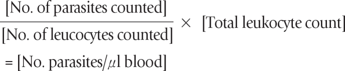
Thin and thick blood films were prepared immediately upon blood collection on the same slide. For thick films, 12 µl of blood was spread over a diameter of 15 mm, while 2 µl of blood was used for thin films. The slides were made in duplicates (one to be read, and the other archived as part of our quality assurance process) and labeled appropriately. The thin film was fixed in absolute methanol for 1-2 sec and air dried. The blood films were stained after 24-48 hr with 3% Giemsa stain solution at pH 7.2. The stained slides were read by 2 competent independent microscopists, Reader 1 and Reader 2. In determining the parasite density, the number of malaria parasites (parasite count) and leukocytes were counted per high power field. The absolute parasite density was calculated using the formula below:
A definitive diagnosis of malaria was made when a reddish chromatin dot with a purple or blue cytoplasm of the malaria parasites are seen together. A slide was pronounced negative when 100 high power fields have been examined using ×100 oil immersion objective lens.
Any discrepant (discordance) result in terms of the presence of parasites is re-read by a more experienced microscopist (Reader 3). The discrepant microscopy result is resolved by calculating the percentage discrepancy. The percentage discrepancy was determined using the formula:
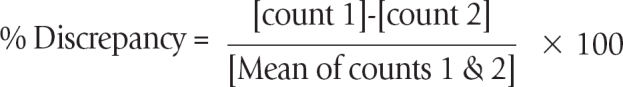
For parasite counts with % discrepancy less than 20%, the count is accepted and the mean parasite count was taken as the parasite density. For parasite counts with % discrepancy ≥ 20%, the films were examined by Reader 3. The count by Reader 3 and the closest from either Reader 1 or Reader 2 were used to calculate the % discrepancy, and their mean count was taken as parasite density provided the % discrepancy is < 20%. Alternatively, the slides were re-read by Reader 1 and Reader 2. The total leukocyte count determination for each participant was done using the improved Neubauer chamber as described by Baker and Silverton [16].
The data were analyzed using EPI INFO 2002 statistical software (CDC, Atlanta, Georgia, USA). Tests for associations and differences were done by chi-square analysis. Statistical significance was set at P value < 0.05.
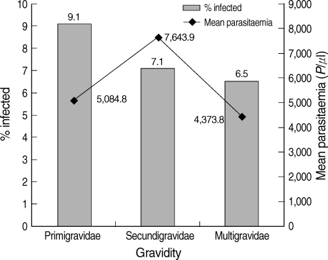
A total of 1,084 pregnant women participated in the study. Their mean age ± SD was 27.4 ± 4.8 year. Of these, 572 (49.8%) were in the second trimester of pregnancy. The pregnant women with secondary education were 42.0%. The characteristics of the participants are shown in Table 1. Positive malaria slides were recorded in 83 pregnant women thus giving a malaria prevalence rate of 7.7% (95% confidence interval; 6.2-9.4%). The parasite density (mean and SD) among the infected pregnant women was 5,420 ± 17,522 parasites/µl. There was a reduction (though not significant, χ2 = 2.06; degree of freedom [df] = 2; P = 0.357) in the proportion of those with malaria infection as gravidity increased, with the primigravidae having the highest, 9.1% (Fig. 1). This association between the age of the pregnant women and proportion of those infected was significant (χ2 = 13.36; df = 4; P = 0.010). The 15-19 year age group had the highest rate of infection (20.5%) (Fig. 2). There was, however, no significant difference (χ2 = 8.51; df = 4; P = 0.075) in the mean parasitemia of the various age groups (Table 2).
Until now, the reports of the prevalence of malaria in pregnancy were variable and high, especially in the South-West Nigeria where prevalence rates of between 36.5% and 72% [2,9,11] have been reported. These reports contrast sharply with our findings in this same region with a prevalence rate of 7.7% among pregnant women attending antenatal clinics for the first time during current pregnancy.
The differences in the reported prevalence rates of malaria may be attributed to the skill and experience of the laboratory personnel involved in blood film preparation, staining, and reading of the slides. In our study, we ensured that the sensitivity and specificity of our microscopists were above 90% through frequent training and the institution of a malaria microscopy quality assurance programme. Strict adherence to procedures for slide preparation and staining [17] ensured the production of clear, well stained slides, thereby reducing errors due to artefacts. The inaccurate diagnosis of malaria is not peculiar to Nigeria. In Tanzania, Mwanziva et al. [18] reported a very appalling case, where < 1% of the slides read by clinic microscopists as malaria positive were confirmed as positive by trained research scientists.
The consequences of over-reporting of malaria cases are: 1) Inability to properly assess the impact of malaria control programmes, as the baseline information before implementation may not be correct; 2) In clinical settings, there will be unnecessary treatments with antimalarial drugs for febrile cases that may not be due to malaria, thereby, increasing the chances of the emergence of antimalarial drug resistance and wastage; 3) National and world malaria statistics based on inaccurate data could misrepresent the actual reality on ground.
Our current results call for a total re-assessment of existing malaria prevalence data. This can only be achieved prospectively after pre-qualifying the microscopists. Our results showed that primigravidas (8.5%) were more often infected than women of other gravidities (Fig. 1). Primigravidas have been reported to be at a greatest risk of malaria in pregnancy because they lack the specific immunity to placental malaria which is acquired from exposure to malaria parasites during pregnancy [4,19]. This immunity accumulates with successive pregnancies, provided there is exposure to malaria infection [20]. The similar malaria prevalence rates in secundigravidas and multigravidas seen in this study suggest that there is no difference in the level of specific immunity to placental malaria and in line with existing data [20]. When this is considered, the reported high prevalence rates of malaria in pregnant women [9-12] seem to nullify the usefulness of this acquired immunity which has been shown to be able to prevent placental malaria by blocking parasite adhesion to placental circumsporozoite antigen (CSA) and opsonize malaria-infected erythrocytes for interaction with Fc receptors on phagocytic cells [19].
In this study, the highest malaria prevalence was seen in pregnant women < 20 year (20.5%) (Fig. 2). This finding is consistent with earlier reports [21,22] where age group of < 24 year was reported to be at a high risk. To stem this trend, awareness on malaria prevention measures during pregnancy should target young women even before they get married preferably at schools, and religious and social gatherings. Our results showed that malaria prevalence decreased with increasing age but increased in age group > 34 year. Marielle et al. [23] reported a high prevalence of malaria in pregnant women within a similar age group (36-39 year) in Gabon.
As our study has shown the over-reporting in the prevalence of malaria in pregnancy, there is need to ensure that competent microscopists whose levels of sensitivity and specificity are critical in settings where laboratory-based confirmation of clinically suspected malarial fever is done. There is, therefore, a need for the institution of a malaria quality assurance programme in laboratories involved in malaria diagnosis.