Warning: mkdir(): Permission denied in /home/virtual/lib/view_data.php on line 81

Warning: fopen(upload/ip_log/ip_log_2024-04.txt): failed to open stream: No such file or directory in /home/virtual/lib/view_data.php on line 83

Warning: fwrite() expects parameter 1 to be resource, boolean given in /home/virtual/lib/view_data.php on line 84
Traumatic Myiasis Caused by an Association of Sarcophaga tibialis (Diptera: Sarcophagidae) and Lucilia sericata (Diptera: Calliphoridae) in a Domestic Cat in Italy
| Home | E-Submission | Sitemap | Contact us |  
top_img
Korean J Parasito Search

CLOSE

Korean J Parasito > Volume 53(4):2015 > Article
Pezzi, Whitmore, Chicca, Lanfredi, and Leis: Traumatic Myiasis Caused by an Association of Sarcophaga tibialis (Diptera: Sarcophagidae) and Lucilia sericata (Diptera: Calliphoridae) in a Domestic Cat in Italy

Abstract

We describe here a rare case of traumatic myiasis occurred in August 2014, caused by an association of 2 Diptera species, Sarcophaga tibialis Macquart (Diptera: Sarcophagidae) and Lucilia sericata (Meigen) (Diptera: Calliphoridae), in a domestic cat in northern Italy. Species identification was based on adult male morphology. The present case is the first report of S. tibialis as an agent of myiasis in Italy, and also the first ever report of myiasis caused by an association of S. tibialis and L. sericata. The cat developed an extensive traumatic myiasis in a large wound on the rump, which was treated pharmacologically and surgically. The biology, ecology, and distribution of S. tibialis and L. sericata are also discussed. A literature review is provided on cases of myiasis caused by S. tibialis, and cases of myiasis by L. sericata involving cats worldwide and humans and animals in Italy.

INTRODUCTION

Myiasis is defined by Zumpt [1] as “the infestation of live human and vertebrate animals with dipterous larvae, which, at least for a certain period, feed on the host’s dead or living tissue, liquid body-substances, or ingested food”. Traumatic myiasis is caused by fly larvae infesting pre-existing traumatic lesions or actively gaining access to tissues [1]. Fly species that cause traumatic myiasis can be divided into obligate or facultative ones according to the nature of the host-parasite association. Obligate traumatic myiasis involves larvae that can only develop on living tissue, whereas in facultative myiasis the larvae can develop on both living and dead tissues [2]. Here, we describe a rare case of traumatic myiasis in a domestic cat (Felis catus L., Mammalia: Felidae) caused by an association of Sarcophaga tibialis Macquart (Diptera: Sarcophagidae) and Lucilia sericata (Meigen) (Diptera: Calliphoridae).

CASE DESCRIPTION

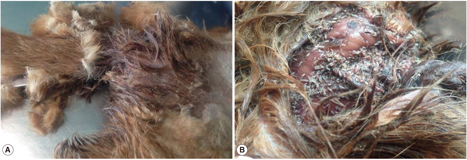
A 10-year old European shorthair cat, missing from home for about 3 days in August 2014, was found near its home in the village of San Martino (Ferrara province, Italy) with a large wound on the rump near the base of its tail, which was almost completely detached (Fig. 1A, B). The cat was brought to a veterinary clinic for treatment. The veterinaries assumed that the injury had been caused by a dog bite and found an extensive traumatic myiasis in the wound caused by a high number of Diptera larvae (Fig. 1B).
The veterinaries treating the cat collected 60 larvae (all first or second instar), which were placed in plastic test tubes and immediately brought to the Laboratory of Urban Ecology, Department of Life Sciences and Biotechnology, University of Ferrara, for identification. The wound was washed with an antiseptic (povidone-iodine), and ivermectin was administered subcutaneously. Afterwards, the cat’s tail was surgically amputated and during surgery the cat was drip-fed with physiological solution, a vitamin support, and a corticosteroid. After suturing the wound the cat was treated with broad-spectrum antibiotics (streptomycin and penicillin).
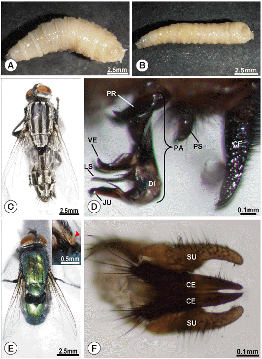
In the Laboratory of Urban Ecology the larvae were divided into 2 groups: 15 of them were immediately fixed in 4% formaldehyde and the other 45 were reared in a plastic box containing 90 g of cow liver, at 25˚C, 50% relative humidity and a 16/8 (L/D) photoperiod. Upon reaching the third instar, the larvae were again divided into 2 groups according to their length (respectively 14.8±0.7 mm and 11.6±1.0 mm) and aspect (Fig. 2A, B) and raised until the adult stage. Morphological investigations were performed with a Nikon SMZ 800 stereomicroscope (Nikon Instruments Europe, Amsterdam, The Netherlands) and a Nikon Eclipse 80i optical microscope (Nikon Instruments Europe), both connected to a Nikon Digital Sight Ds-Fil camera (Nikon Instruments Europe).
The identification of S. tibialis was carried out by examination of adult males (Fig. 2C) emerged from the first group of larvae, based mainly on aspects of the genitalia (Fig. 2D) and using the identification key of Richet et al. [3]. The identification of the second species as L. sericata was carried out using the identification key of Szpila [4] and was based on morphology of adult males (Fig. 2E) emerged from the second group of larvae, namely the bright yellow basicosta (Fig. 2E, inlay) and shape of the cerci and surstyli (Fig. 2F). All examined larvae and adults are deposited in the Laboratory of Urban Ecology, Department of Life Sciences and Biotechnology, University of Ferrara, Ferrara (Italy).

DISCUSSION

Reports of myiasis in cats are uncommon, probably because they usually groom themselves carefully [5]. In the present case, infestation by these 2 fly species was favoured by a deep and extensive wound that had weakened the cat, preventing it from properly grooming and cleaning its fur. Cases of myiasis in humans and animals caused by an association of more than one Diptera species have been reported in the literature. The species involved are Chrysomya rufifacies (Macquart) (Diptera: Calliphoridae) with Chrysomya megacephala (Fabricius) (Diptera: Calliphoridae) [6], Sarcophaga ruficornis (Fabricius) (Diptera: Sarcophagidae) with Musca domestica (Linnaeus) (Diptera: Muscidae), and C. megacephala [7] and Chrysomya bezziana Villeneuve (Diptera: Calliphoridae) with Lucilia sp. (Diptera: Calliphoridae) [8].
Myiasis caused by S. tibialis in association with another species had been reported only once, by Villeneuve [9] with Sarcophaga crassipalpis Macquart (Diptera: Sarcophagidae). L. sericata has been reported to cause myiasis in association with Chrysomya albiceps Wiedemann (Diptera: Calliphoridae) [5] and Wohlfahrtia magnifica (Schiner) (Diptera: Sarcophagidae) [10,11]. The case we report is the first of myiasis caused by an association of S. tibialis and L. sericata.
S. tibialis is a thermophilic, heliophilic, and generally synanthropic species [12], probably indigenous to the Afrotropical Region [13] and widespread in Africa (including the Canary Islands, Madagascar, and the Seychelles), central and southern Europe, Turkey, the Chagos Archipelago in the Oriental Region, and Oceania [14]. The species is widespread in Italy, including Sardinia and Sicily [15]. Like most Sarcophagidae, S. tibialis is larviparous [16], with eggs usually hatching inside the uterus. Eggs laid in groups or together with larvae have been reported not to hatch [17]. As in most flies, the rate of development is largely temperature-dependent [1]. Abasa [17] reported that at 20-22˚C the development from first larval stage to adult lasts about 20-27 days, whereas Aspoas [18] stated that at 25˚C development from first instar to adult lasts 20-22 days. Recently, Villet et al. [19] reported that the optimal temperature for development of this species (based on lowest larval mortality) is about 20˚C.
In our study, females of S. tibialis and L. sericata were attracted by the smell released by the wound and subsequently deposited larvae and eggs on it. Adults of S. tibialis are usually attracted by decaying matter such as carcasses and excrements [13,17,20], where larvae may or may not be laid. Larvae of this species have been reared in the laboratory on liver, fish carrion, fresh bird meat, dead snails, and chopped grasshoppers [3,17-19]. S. tibialis is also of potential forensic importance [12].
Only 2 well-documented cases of cutaneous human myiasis caused by S. tibialis were reported in the literature. Both occurred in Tripoli (Libya) in 1913, the first in May and the second in July. Both cases were connected to poor sanitation and the myiasis was localized to the scalp. The first case involved a bedridden boy affected by peritoneal tuberculosis and tinea capitis [16], the second occurred in a little girl affected by tinea capitis, pediculosis, and pyoderma [21]. According to these reports, the smell released by the infected scalp lesions attracted the adult flies. Onorato [16] hypothesized a possible connection between tinea capitis and cutanous myiasis by S. tibialis. Besides these 2 cases, there is a hint of another case of human traumatic myiasis in Algeria mentioned by Villeneuve [9], but without any further detail. Also, Patton and Evans [22] stated that S. tibialis is known to cause intestinal myiasis in Europe, but they did not report any data on the number of cases and localities of occurrence. Therefore, the present case is the first record of S. tibialis causing myiasis in Italy and is probably the first well-documented case of this kind in Europe.
On the other hand, L. sericata is a widely distributed agent of myiasis in temperate areas [2] and a probable cosmopolite [23]. In the United Kingdom, South Africa, and New Zealand, L. sericata is a relevant agent of myiasis in sheep [24]. It is widespread in the whole of Italy, including Sardinia and Sicily [15,25]. Adults are diurnal and commonly breed on carcasses, dirty or wet sheep fleece, garbage and manure; they are also attracted by open wounds and lesions where conditions are such that the larvae can complete their development, causing myiases such as fly strike in sheep. Females start to lay eggs 5-9 days after emerging from the puparium and within 3 weeks they lay 2,000-3,000 eggs in 9-10 batches [1]. The larval developmental rate is strongly temperature-dependent: between 22˚C and 29˚C, development from egg to adult occurs in 13-19 days [23].
In southern Europe, L. sericata is considered a synanthropic species associated to houses and rural areas undergoing urbanization [26,27]. Together with Calliphora vicina Robineau-Desvoidy (Diptera: Calliphoridae) and Lucilia illustris Meigen (Diptera: Calliphoridae), L. sericata is among the 3 species characterizing urban and suburban areas in Europe [28]. In our report the cat contracted myiasis in San Martino, a small country village about 4 km from the city of Ferrara (Ferrara province, Italy). Myiasis caused by L. sericata has been reported in domestic cats in America, Europe, and Asia and may be traumatic [5,29-33], gastrointestinal (anal and rectal) [5,34], urogenital (vaginal, penile and perineal) [30,35-37], or ocular [38]. Our report is the first case of traumatic myiasis by L. sericata in a domestic cat in Italy, albeit in association with S. tibialis. The only other case of myiasis by L. sericata in a domestic cat in Italy was a rectal one, although cutaneous myiases by this species have been reported in sheep and dogs [34], and humans [39,40].
The present results on the first case of myiasis caused by an association of S. tibialis and L. sericata and on the first case of myiasis by S. tibialis in Italy were obtained through a close cooperation between veterinaries and entomologists. Studies on the biology, distribution, and modes of infestation of Diptera causing myiasis could largely benefit from similar collaborations. Accurate and detailed information on the distribution of species of medical and veterinary interest is limited in Italy [41]. Thus, the compilation of an infestation map should be a priority objective for veterinary interventions and for ecological and biological studies.

ACKNOWLEDGEMENTS

We owe thanks to Dr. Cecilia Farinella and Dr. Silvia Cazzola, the veterinaries who signalled the case. We also wish to thank Mr. Nigel Wyatt (Natural History Museum, London, UK) for confirming the identification of Lucilia sericata, and Dr. Massimo Tassinari (Animal Health Sector, Azienda USL, Ferrara, Italy) for critically reviewing the manuscript and providing relevant clinical suggestions. Funds supporting this study were provided by CTB-PROG-ZT-2012, AUSL Cesena, assigned to the Department of Life Sciences and Biotechnology, University of Ferrara (Ferrara, Italy). The manuscript is dedicated to the loving memory of Mr. Domenico Bardi.

Conflict of interest

The authors declare no conflict of interest related this work.

REFERENCES

1. Zumpt F. Myiasis in man and animals in the old world. London, UK. Butterworth & Co. 1965, pp 1-107.

2. Hall MJR, Farkas R. Traumatic myiasis of humans and animals. In Papp L, Darvas B eds, Contributions to a Manual of Palaearctic Diptera (with special reference to flies of economic importance), Vol. 1. General and Applied Dipterology. Budapest, Hungary. Science Herald. 2000, pp 751-768.

3. Richet R, Blackith RM, Pape T. Sarcophaga of France (Diptera: Sarcophagidae). Sofia, Bulgaria. Pensoft Publishers. 2011, pp 5-327.

4. Szpila K. Key for identification of European and Mediterranean blowflies (Diptera, Calliphoridae) of medical and veterinary importance - adult flies. In Gennard D ed, Forensic Entomology, An Introduction. 2nd ed. West Sussex, UK. Wiley-Blackwell. 2012, pp 77-81.

5. Schnur HJ, Zivotofsky D, Wilamowski A. Myiasis in domestic animals in Israel. Vet Parasitol 2009;161:352-355.
crossref pmid
6. Sukontason KL, Narongchai P, Sripakdee D, Boonchu N, Chaiwong T, Ngern-Klun R, Piangjai S, Sukontason K. First report of human myiasis caused by Chrysomya megacephala and Chrysomya rufifacies (Diptera: Calliphoridae) in Thailand, and its implication in forensic entomology. J Med Entomol 2005;42:702-704.
crossref pmid
7. Ferraz ACP, Proença B, Gadelha BQ, Faria LM, Barbalho MGM, Aguiar-Coelho VM, Lessa CSS. First record of human myiasis caused by association of the species Chrysomya megacephala (Diptera: Calliphioridae), Sarcophaga (Liopygia) ruficornis (Diptera: Sarcophagidae), and Musca domestica (Diptera: Muscidae). J Med Entomol 2010;47:487-490.
crossref pmid
8. Obanda V, Ndambiri EM, Kingori E, Gakuya F, Lwande OW, Alasaad S. Traumatic myiasis in free-ranging eland, reported from Kenya. Parasit Vectors 2013;6:89.
crossref pmid pmc
9. Villeneuve J. Sur les larves de Sarcophaga. Bull Soc Entomol Belgique 1922;4:91-92.

10. Seif AI, Kholeif HA, Koura EA, Alfi NM. Experience with traumatic myiasis among patients with septic wounds. J Trop Med 1992;2:115-119.

11. Salimi M, Goodarzi D, Karimfar MH, Edalat H. Human urogenital myiasis caused by Lucilia sericata (Diptera: Calliphoridae) and Wohlfahrtia magnifica (Diptera: Sarcophagidae) in Markazi Province of Iran. Iran J Arthropod Borne Dis 2010;4:72-76.
pmid pmc
12. Povolný D, Verves YG. The flesh-flies of Central Europe (Insecta, Diptera, Sarcophagidae). Spixiana Supplement 1997;24:1-260.

13. Zumpt F, Patterson PM. Flies visiting human faeces and carcasses in Johannesburg, Transvaal. South Afr J Clin Sci 1952;3:92-106.

14. Pape T. Catalogue of the Sarcophagidae of the World (Insecta: Diptera). Mem Entomol Int 1996;8:1-558.

15. Pape T, Richter V, Rivosecchi L, Rognes K. Diptera Hippoboscoidea, Oestroidea. In Minelli A, Ruffo S, La Posta S eds, hecklist delle specie della fauna italiana, fascicolo 78. Bologna, Italy. Calderini. 1995, pp 1-36.

16. Onorato R. Le miasi in Tripolitania. Arch Ital Sci Med Colon 1922;3:14-315.

17. Abasa RO. Reproductive biology of Sarcophaga tibialis (Diptera: Sarcophagidae). I. Life history with notes on prepupation mortality and pupation habits. Ann Entomol Soc Am 1970;63:466-469.
crossref
18. Aspoas BR. Comparative micromorphology of third instar larvae and the breeding biology of some Afrotropical Sarcophaga (Diptera: Sarcophagidae). Med Vet Entomol 1991;5:437-445.
crossref pmid
19. Villet MH, MacKenzie B, Muller WJ. Larval development of the carrion-breeding flesh fly, Sarcophaga (Liosarcophaga) tibialis Macquart (Diptera: Sarcophagidae), at constant temperatures. Afr Entomol 2006;14:357-366.

20. Aspoas BR. Afrotropical Sarcophagidae in a carrion fly community. Med Vet Entomol 1994;8:292-294.
crossref pmid
21. De Napoli F. Sopra un caso di dermato-myiasis muscosa da Sarcophaga beckeri osservato a Tripoli. Ann Med Nav e Col 1914;20:343-358.

22. Patton WS, Evans AM. Insects, ticks, mites and venomous animals of medical and veterinary importance. Part I. Medical. Croydon, UK. HC Grubb Ltd. 1929, pp 492-493.

23. Robinson WH. Urban insects and arachnids: a handbook of urban entomology. Cambridge, UK. Cambridge University Press. 2005, p 154.

24. Spradbery JP. A manual for the diagnosis of screw-worm fly. Agriculture Fisheries and Forestry, Commonwealth of Australia. Canberra, Australia. 2002, pp 17-18.

25. Rognes K. Short notes 12. Diptera, Calliphoridae. In Nardi G, Whitmore D, Bardiani M, Birtele D, Mason F, Spada L, Cerretti P eds, Biodiversity of Marganai and Montimannu (Sardinia). Research in the framework of the ICP Forests network. Conservazione Habitat Invertebrati, 5. Sommacampagna (Verona), Italy. Cierre Edizioni. 2011, pp 852-857.

26. Turchetto M, Lafisca S, Costantini G. Postmortem interval (PMI) determined by study sarcophagous biocenoses: three cases from the province of Venice (Italy). Forensic Sci Int 2001;120:28-31.
crossref pmid
27. Vanin S, Tasinato P, Ducolin G, Terranova C, Zancaner S, Montisci M, Ferrara SD, Turchetto M. Use of Lucilia species for forensic investigations in Southern Europe. Forensic Sci Int 2008;177:37-41.
crossref pmid
28. Hwang C, Turner BD. Spatial and temporal variability of necrophagous Diptera from urban to rural areas. Med Vet Entomol 2005;19:379-391.
crossref pmid
29. Ribbeck R, Schröder E, Schumann H. Lucilia-sericata-Larven als Erreger von Wundmyiasis bei Hund und Katze. Mh Vet-Med 1979;34:383-384.

30. Hinaidy HK, Frey H. Weitere Fakultativmyiasis-Fälle bei Wirbeltieren in Österreich. Wien Tierärztl Monat 1984;71:237-238.

31. Mariluis JC, Schnack JA, Cerverizzo I, Quintana C. Cochliomyia hominivorax (Coquerel, 1858) and Phaenicia sericata (Meigen, 1826) parasiting domestic animals in Buenos Aires and vicinities (Diptera, Calliphoridae). Mem Inst Oswaldo Cruz 1994;89:139.
crossref pmid
32. Vignau ML, Arias DO. Myiasis cutáneo-ulcerosas en pequeños animales. Parasitol Día 1997;21:36-39.

33. Dik B, Uslu U, Işik N. Myiasis in animals and human beings in Turkey. Kafkas Univ Vet Fak Derg 2012;18:37-42.

34. Principato M, Cioffi A. Notes on the incidence of the Lucilia genus (Diptera: Calliphoridae) in Umbria, Central Italy. A case of myiasis by Lucilia ampullacea (Villen 1922) in Testudo graeca. In 20t International Congress of Entomology. 25-31 August 1996; Florence, Italy. Proceedings: 1996. p. 769.

35. Hall RD. The blow flies of Missouri: an annotated checklist (Diptera: Calliphoridae). Trans Mo Acad Sci 1979;13:33-36.

36. Yücel Ş, Çiçek H, Kar S, Eser M. Bir kedide genital myiasis olgusu. Türkiye Parazitol Derg 2008;32:241-243.
pmid
37. Yilmaz O, Kose M. Vulvo-vaginal myiasis in a queen caused by Lucilia sericata (Diptera: Nematocera: Calliphoridae). Eurasian J Vet Sci 2014;30:108-110.

38. Eren H, Aypak S, Ural K, Seven F. Traumatic myiasis in a dog and ocular myiasis in a cat cases due to Lucilia sericata (Diptera: Calliphoridae) larvaes. Kafkas Univ Vet Fak Derg 2010;16:883-886.

39. Majocchi D. Sopra due nuovi casi di dermato-myiasis muscosa da Lucilia sericata. Memorie R Accad Sci Ist Cl Sci fis Bologna 1920;7:237-253.

40. Dutto M, Pomero F, Migliore E, Fenoglio L. Cutaneous myiasis in a geriatric patient. Parassitologia 2010;52:435-438.
pmid
41. Bonacci T, Greco S, Whitmore D, Curcio U. First data on myiasis caused by Wohlfahrtia magnifica (Schiner, 1862) (Insecta: Diptera: Sarcophagidae) in Calabria, southern Italy. Life Excit Biol 2013;1:197-201.
crossref

Fig. 1.
Clinical aspects of a case of traumatic myiasis caused by Sarcophaga tibialis and Lucilia sericata. (A) Wound on the cat’s rump, near the base of the tail. (B) Wound detail showing the fly larvae.
kjp-53-4-471f1.gif
Fig. 2.
Morphology of Sarcophaga tibialis and Lucilia sericata. (A) Third-instar larva of S. tibialis. (B) Third-instar larva of L. sericata. (C) Habitus in dorsal view of adult male of S. tibialis. (D) Detail of male genitalia of S. tibialis. (E) Habitus in dorsal view of adult male of L. sericata; the inlay shows the base of the left wing in dorsal view and the arrow indicates the yellow basicosta. (F) Dorsal view of cerci and surstyli of L. sericata. Terminology follows Richet et al. [3] and Szpila [4]. CE, cercus; DI, distiphallus; JU, juxta; LS, lateral stylus; PA, phallus; PR, pregonite; PS, postgonite; VE, vesica; SU, surstylus.
kjp-53-4-471f2.gif
Editorial Office
Department of Molecular Parasitology, Samsung Medical Center, School of Medicine, Sungkyunkwan University,
2066 Seobu-ro, Jangan-gu, Suwon 16419, Gyeonggi-do, Korea.
Tel: +82-31-299-6251   FAX: +82-1-299-6269   E-mail: kjp.editor@gmail.com
About |  Browse Articles |  Current Issue |  For Authors and Reviewers
Copyright © 2024 by The Korean Society for Parasitology and Tropical Medicine.     Developed in M2PI