INTRDUCTION
Digenetic trematodes are a relatively large trematode group comprising of more than 2,500 nominal genera. It commonly has 3 hosts (2 intermediate and 1 definitive) in the life cycle. Various kind of animals, i.e., mollusc, fish, amphibian, reptilian, and arthropod, act as the second intermediate hosts, which retained the infective larvae, i.e., metacercariae. Especially, it has been known that some species of digenetic trematode metacercariae are detected in molluscan hosts, the clam and oyster [1–3]. In the Republic of Korea (=Korea), some species of gymnophallid metacercariae were detected from the intertidal clams and oysters [4–11]. Some species of echinostomatid metacercariae were also found in the brackish water gastropods [12–16].
Yu et al. [4] first detected Parvatrema duboisi (syn. P. timondavidi) metacercariae from Manila clams, Ruditapes philippinarum, and Sohn et al. [10] recorded Parvatrema chaii n. sp. (Digenea: Gymnophallidae), which were recovered from mice experimentally infected with the metacercariae collected from the surf-clam Mactra veneriformis. Lee et al. [5] first found that the oyster, Crassostrea gigas, is the second intermediate host (the source of human infection) of Gymnophalloides seoi. Kim and Yun [9] surveyed on larval trematodes in 3 species of bivalves, Corbicula japonica, Sinonovacula constricta, and R. philippinarum, from some sites in the Yellow Sea. Chai et al. [11] reported a new gymnophallid trematode, Meiogymnophallus sinonovaculae n. sp., recovered from the small intestines of mice infected with metacercariae from the razor clam, S. constricta. On the other hand, Kim and Chun [12] detected Himasthla kusasigi metacercariae in the clam, Meretrix lusori. Kim [13] also found 2 species of echinostomatid metacerceriae, H. kusasigi and Acanthoparyphium tyosenense, from 3 species of intertidal clams, i.e., M. veneriformis, Cyclina sinensis, and Solen strictus. After then, Chai et al. [14] discovered human infections with A. tyosenense, and they also investigated the metacercarial infections of this fluke in 5 species of bivalves, i.e., M. veneriformis, Solen grandis, Meretrix petechialis, C. sinensis, and Scapharca broughtonii, and a marine snail, Neverita bicolor, to survey on the sources of human infections. Han and Chai [15] reported for the first time that M. veneriformis act as the second intermediate host of Acanthoparyphium marilae.
The infection status of Parvatrema sp. metacercariae was investigated in R. philippinarum collected from southern coastal areas of Korea [6]. Lee et al. [7] performed a nationwide survey of naturally produced oysters to know the infection status with G. seoi metacercariae. Most of the previous studies were performed to identify the specific trematode species from the limited host animals and areas [4–16]. However, the metacercarial infection status of intertidal bivalves collected from the western coastal regions has not been widely and systematically examined. Therefore, we investigated the infection status of 5 species of bivalves from 4 local sites in the western coast of Korea with the trematode metacercariae.
MATERIALS AND METHODS
Subjected samples by surveyed areas
In July 2013, total 4 species of clams (Mactra veneriformis, Ruditapes philippinarum, Cyclina sinensis and Saxidomus purpuratus) were collected from 3 western coastal regions (Fig. 1), i.e., Taean-gun (latitude: 36.4710; longitude: 126.3408) in Chungcheongnam-do, Buan-gun (35.0727; 126.0734) and Gochang-gun (35.5009; 126.4907) in Jeollabuk-do, Korea. A total of 90 M. veneriformis were collected in all 3 regions, Taean-gun (n=30), Buan-gun (n=20), and Gochang-gun (n=40). Two species of clams, R. philippinarum (n=25) and C. sinensis (n=25), were also collected with the same number in 2 regions, Taean-gun (n=15) and Gochang-gun (n=10), respectively. In Buan-gun, 10 S. purpuratus were collected. To investigate the recent infection status of G. seoi metacercariae, oysters, Crassostrea gigas, were collected in July 2013 from a tidal flat of Aphae-myeon (Township) in Shinan-gun (34.0839; 126. 3508), Jeollanam-do, a well-known endemic area of this fluke [16].
Examination of the metacercarial infection status in clams and oysters
All collected clams and oysters were transferred on ice to the laboratory of the Department of Parasitology and Tropical Medicine, Gyeongsang National University School of Medicine, Jinju, Korea. After species identification, the bivalves were individually opened with a knife. Each opened sample (the animal part and 2 shells) was mixed with artificial gastric juice, and the mixture was incubated at 36°C for 1–2 hr with occasional stirring (at 15–20 min interval). The digested material was filtered through a 1×1 mm mesh, and washed with 0.85% saline until the supernatant became clear. The sediment was carefully examined under a stereomicroscope. Each species of metacercariae were separately collected by the general feature, and they were counted to get hold of the infection rate (%) and density (no. of metacercariae per clam infected) by clam species.
Observation of metacercariae and experimental infection to animals
Some representative metacercariae collected with a stereomicroscope were morphologically observed under a light microscope equipped with a micrometer (OSM-4, Olympus, Tokyo, Japan) to identify their exact species. Especially, the metacercariae of Parvatrema spp. detected in M. veneriformis from Taean-gun were fixed with 10% formalin under a cover slip, and then stained with Semichon’s acetocarmine to observe their morphological characteristics. The collected metacercariae were orally infected with a gavage needle to chicks and hamster to obtain adult worms. Two species of echinostomes, Himasthla alincia and Acanthoparyphium tyosenense, were recovered in the small intestines of chicks at 2 weeks after infection. Many specimens of Parvatrema spp. were also recovered in the small intestines of hamsters at day 5 after the infection. The experimental animals, hamsters and chicks, were treated according to the guidelines of Institutional Animal Care and Use Committee (IACUC) in Gyeongsang National University. The recovered worms were fixed with 10% formalin under a cover glass pressure, and then stained with Semichon’s acetocarmine to observe their morphological characteristics.
RESULTS
Infection status of M. veneriformis with trematode metacercariae
More than 3 species of metacercariae, i.e., Himasthla alincia, A. tyosenense, and Parvatrema spp., were detected in M. veneriformis. The metacercariae of H. alincia were found in 98.9% clams, and their mean density was 30.8 per clam infected. The metacercariae of A. tyosenense were detected in 47.8% clams, and their mean density was 2.5 per clam infected. Parvatrema spp. metacercariae were found in 43.3% clams, and their mean density was 2,018 per clam infected. The metacercarial infection status by the surveyed area is designated in detail in Table 1.
Infection status of R. philippinarum with trematode metacercariae
More than 2 species of metacercariae, i.e., H. alincia and Parvatrema spp., were detected from R. philippinarum. The metacercariae of H. alincia were found in 80.0% and 30.0% clams from Taean-gun and Gochang-gun, and their mean densities were 5.8 and 1.7 per clam infected, respectively. Parvatrema spp. metacercariae were detected in 6.7% and 100% clams from Taean-gun and Gochang-gun, and their mean densities were 126 and 238 per clam infected, respectively (Table 2).
Infection status of C. sinensis and S. purpuratus with trematode metacercariae
Only 1 species, H. alincia, was detected from C. sinensis clams. The metacercariae were found in 93.3% and 100% clams from Taean-gun and Gochang-gun, and their mean densities were 5.5 and 60.5 per clam infected, respectively (Table 3). No metacercariae were found in 10 S. purpuratus collected in Buan-gun, Jeollabuk-do.
Infection status of oysters with G. seoi metacercariae
The metacercariae of G. seoi were detected in all 30 oysters (100%) collected from Aphae-myeon in Shinan-gun, Jeollanam-do, and their density was 646 per oyster in average. The metacercarial density by the weight group of oysters is shown in detail in Table 4.
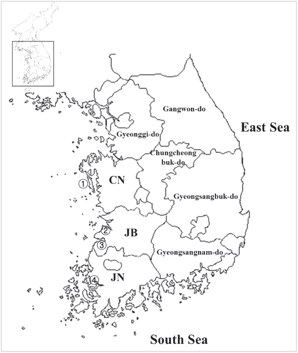
Morphology of H. alincia metacercariae and adults
Metacercariae (Fig. 2A): Cyst oval or round, 240–257 (249) x232–250 (242) in size with 5–9 (7) thick cyst wall, had a head collar with 31 collar spines that included 4 lateral end-group spines on each side, and 2 excretory ducts filled with numerous fine granules.
Adults (Fig. 2B): Body markedly elongate, maximum width at the level of the anterior testis. Head crown distinct, bearing 31 collar spines arrange in a single uninterrupted row, with 4 end group spines on each side of ventral corners (Fig. 2C). Oral sucker small, subterminal. Prepharynx very short. Pharynx subglobular. Esophagus short. Ceca bifurcating near the anterior margin of the ventral sucker, terminating blindly near the posterior extremity. Ventral sucker slightly elliptical. Sucker ratio (oral sucker:ventral sucker) approximately 1:2.93. Cirrus sac elongate, dorsal to ventral sucker, containing seminal vesicle, prostate gland cells, and a cirrus. Ovary spherical, on the median or slightly dextral. Mehlis’ gland located between the ovary and the anterior margin of the anterior testis. Two testes oval to elliptical, tandem, intercecal, near the posterior end of the body. Vitellaria follicular, distributing laterally from anterior 1/3 of the body to near the posterior end. Uterus with eggs intercecal, from the Mehlis’ gland to the anterior 2/3 of the body. Eggs operculate, golden yellow, 93–108 (100) by 55–63 (60). Dimensions of each organ are revealed in detail in Table 5.
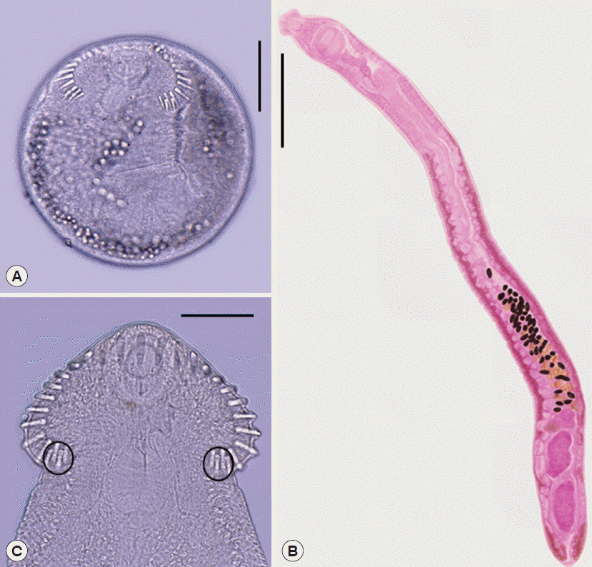
Morphology of Acanthoparyphium tyosenense metacercariae and adults
Metacercariae (Fig. 3A): Cyst oval to round, 350–385 (377) in diameter, with thin cyst wall, a coiled larva and extensively distributed excretory granules. Two suckers, oral and ventral sucker, plainly visible. Head collar reniform, with 23 collar spines in a single row, without ventral corner spines.
Adults (Fig. 3B): Body elongate, medium, maximum width in acetabula level. Head crown distinct, bearing about 23 collar spines in a single row, without end group ones (Fig. 3C). Oral sucker small, subterminal. Prepharynx absent. Pharynx subglobular. Esophagus short. Ventral sucker large, well developed. Sucker ratio (oral sucker:ventral sucker) approximately 1:2.93. Cirrus sac well developed and contained saccular seminal vesicle. Ovary spherical, on the dextro-median line of the body. Two testes spherical, tandem. Vitellaria follicular, distributing laterally from the level of anterior testis to posterior end. Eggs operculate, golden yellow. Dimensions of each organ are shown in detail in Table 6.
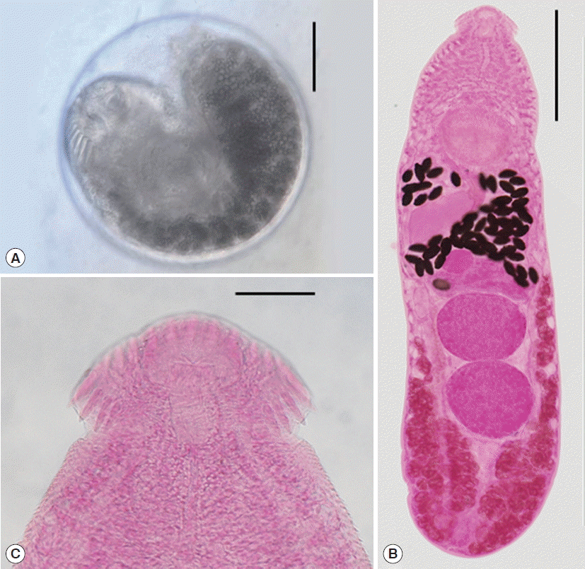
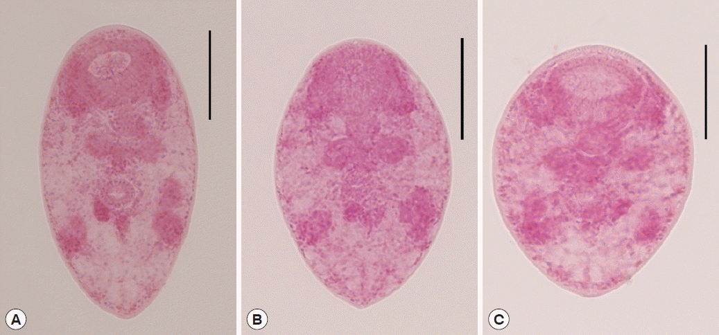
Morphology of Parvatrema spp. metacercariae collected in M. veneriformis from Taean-gun
The metacercariae of Parvatrema spp. collected in M. veneriformis from Taean-gun, Chungcheongnam-do were morphologically divided into 3 types (Fig. 4). Generally, their body very tiny and ovoid, with fine tegumental spines, maximum width at acetabular level. Oral sucker large and well developed, with lateral projections on each side. Prepharynx absent. Pharynx round and muscular. Ceca inflated oval sacs, extended to the level of the ventral sucker. Ventral sucker round, posterior to midline. Sucker ratio was 2.49:1 in average. Ovary oval to round and slightly smaller than testes, dextroanterior to the right testis. Testes round, nearly symmetrical, posterolateral to the ventral sucker. Genital pore, a wide slit-like opening, in the middle between the oral and ventral suckers. Excretory bladder V-shaped with 2 arms extending to the oral sucker level. Dimensions of each organ by the morphological type are designated in detail in Table 7.
DISCUSSION
By the present study, it has been clarified that more than 3 species of trematode metacercariae, i.e., H. alincia, A. tyosenense, and Parvatrema spp., are prevalent in 3 species of clams (M. veneriformis, R. philippinarum, and C. sinensis) from 3 west coastal regions, i.e., Taean-gun, Buan-gun, and Gochang-gun. G. seoi metacercariae were still prevalent in oysters from Shinan-gun, Jeollanam-do. Among more than 4 species of trematodes detected in this study, at least 2 (A. tyosenense and G. seoi) are human-infecting flukes [17–20]. Chai et al. [14] first reported 10 human cases of A. tyosenense infections, with 1–107 worm burdens, in an endemic area of heterophyid flukes in Buan-gun, Jeollabuk-do, Korea [21]. Lee et al. [17] described Gymnophalloides seoi n. sp. (Digenea: Gymnophallidae), which was recovered from a Korean woman suffering from acute pancreatitis and gastrointestinal troubles. This human case was the first human infection by a gymnophallid [17]. After then, the village where the patient resided was found to be a high endemic area of G. seoi; additional endemic areas were also found thereafter [18–22].
Chai et al. [14] investigated 5 species of bivalves (M. veneriformis, S. grandis, M. petechialis, C. sinensis, and S. broughtonii) and 1 species of marine snail (N. bicolor) to know the infection sources of A. tyosenense. They detected A. tyosenense metacercariae in 3 molluscan species, M. veneriformis, S. grandis, and N. bicolor, purchased from the endemic area (Buan-gun, Jeollabuk-do). The prevalence of A. tyosenense metacercariae was 100% in 3 molluscan species, and the density was 134 metacercariae in M. veneriformis, 133 in S. grandis, and 23 in N. bicolor. Kim [13] also surveyed the metacercarial infection status in 3 species of clams, M. veneriformis, C. sinensis, and Solon strictus, collected from Naechodo (Island) in the estuary of Geumgang (River); total 208 A. tyosenense metacercariae were detected in 63 out of 100 S. strictus examined. In the present study, A. tyosenense metacercariae were found only in M. veneriformis collected from Taean-gun and Gochang-gun, and their prevalences (mean density/clam infected) were 50.0% (2.1) in Taean-gun and 70.0% (2.8) in Gochang-gun. Unlike Chai et al. [14], A. tyosenense metacercariae were not detected in 20 M. veneriformis collected in Buan-gun in this study. The endemicity of A. tyosenense metacercariae in this study was more or less similar with that of Kim [13]; however, it was very low when compared with that of Chai et al. [14].
Since Lee et al. [5] first reported that the oyster is the second intermediate host of G. seoi, some epidemiological studies on the infection status of oysters with metacercariae were done in coastal areas of Korea [5,7,8]. Especially, the oysters naturally produced in Aphae-myeon, Shinan-gun, Jeollanam-do [18] were most frequently investigated. Lee et al. [5] detected 2–4,792 (av. 610) metacercariae of G. seoi in 50 oysters from Aphae-myeon. Later, Lee et al. [7] also reported av. 786 metacercariae in 20 oysters from Aphae-myeon. Sohn et al. [8] investigated the monthly infection status with G. seoi metacercariae in 20 oysters each time collected in the same site of Aphae-myeon; total 248 (88.6%) oysters were infected with av. 1,339 metacercariae. In the present study, G. seoi metacercariae were detected in all 30 (100%) oysters from Aphae-myeon, with the mean density of 646 per oyster. Therefore, the mean metacercarial density in this study was similar with that of Lee et al. [5], slightly lower than that of Lee et al. [7], and much lower than that of Sohn et al. [8]. On the other hand, Lee et al. [5] mentioned that the metacercarial density had no relationship with the size of oysters. However, in Sohn et al. [8] and the present study, the metacercarial density was proportional to the increase of the oyster weight.
Among the 27-nominated species of the genus Himasthla, 3 species, namely, H. alincia, H. kusasigi, and H. megacotyle, were reported in Korea [12,16,23]. The morphological characteristics of H. alincia are highly similar to those of H. kusasigi, especially in that they commonly have 31 collar spines, whereas H. megacotyle bears 28 collar spines. However, H. kusasigi is distinguished from H. alincia in that it has a more slender body and its vitelline follicles extended more anteriorly toward the end of the cirrus sac (Table 7) [24,25]. In this respect, our specimens were identified as H. alincia (Fig. 1). The metacercariae of this fluke were detected in 5 species of bivalves (M. veneriformis, S. grandis, C. sinensis, M. petechialis, and R. philippinarum) from a coastal area in Buan-gun, Jeollabuk-do [16]. In the present study, they were detected in 3 species of bivalves (M. veneriformis, C. sinensis, and R. philippinarum) from 3 western coastal areas (Taean, Buan, and Gochang) of Korea. The metacercarial density was much higher in Han et al. [16] than in this study.
In the genus Acanthoparyphium, 14 species, namely, A. phoenicopteri, A. spinulosum, A. ochthodromi, A. marilae, A. squatarolae, A. charadrii, A. kurogamo, A. melanittae, A. spinulosum suzugamo, A. tyosenense, A. paracharadrii, A. loborchis, A. haematopium, and A. macracanthum, have been listed so far [26–28]. Of these, A. tyosenense was originally described with worms from the small intestine of the duck Melanitta fusca stejnegeri and M. nigra americana caught in Korea [24]. This fluke was treated as a synonym of A. kurogamo by Skrjabin [29] and Chen [27]. However, A. tyosenense is a valid species, and it differs from A. kurogamo by its body shape and position of the acetabulum and testes [14,24]. Although another species, A. marilae, has been reported in Korea [15], the morphological characteristics of our specimens are well corresponded with those of A. tyosenense (Fig. 2). Comparative dimensions are designated in Table 6.
Until now, 3 species in the genus Parvatrema (Digenea: Gymnophallidae), i.e., P. duboisi (=P. timondavidi), P. chaii, and P. homoeotecnum, have been reported in Korea [4,10,23]. Among them, P. homoeotecnum was described only with adults, which were detected in the intestines of a migratory bird, the Mongolian plover (Charadrius mongolus), from the coastal area of Gunsan-si (City), Jeollabuk-do [23]. The remaining 2 species were described not only based on the adults but also on the metacercarial stage. Yu et al. [4] first detected P. duboisi metacercariae from intertidal clams, R. philippinarum, and described with adults recovered from experimental mice. Sohn et al. [6] subsequently surveyed on the infection status with Parvatrema sp. metacercariae in R. philippinarum from 13 areas in southern coastal areas of Korea. After that time, Sohn et al. [10] erected a new species, P. chaii n. sp., with adults recovered from mice experimentally infected with metacercariae from surf-clams, M. veneriformis, which were collected from a tidal flat located in Seocheon-gun, Chungcheongnam-do, Korea. In the present study, we found Parvatrema spp. metacercariae in 2 species of clams, R. philippinarum and M. veneriformis. Especially, in M. veneriformis clams collected from Taean-gun, lots of Parvatrema spp. metacercariae were detected and they were morphologically divided into 3 types (Fig. 3). The metacercariae Type A were morphologically similar to P. chaii metacercariae although their body shape (ratio of BL/BW=1.76:1) was slightly different from that of P. chaii (1.98:1). The ratio of OS/VS was the same, 2.60:1, and the ratio of OS/BL was slightly different from each other (Table 7). Moreover, P. chaii metacercariae were previously detected in the same clam hosts, M. veneriformis, collected in Seocheon-gun, Chungcheongnam-do, which is located nearby in the surveyed area of this study, Taean-gun [10]. However, the remaining 2 types of metacercariae (Type B and C) were morphologically different from P. chaii metacercariae [10]. The metacercariae Type B was apparently similar to P. duboisi metacercariae; however, their body size was much smaller than that of P. duboisi. Further studies with molecular analysis are needed in the future to clarify the taxonomic positions of Parvatrema spp. in Korea.