Trematode members in the genus Euryhelmis (Digenea: Heterophyidae) are small, rectangular or pyriform intestinal parasites of frog- and salamander eating mammals [1,2]. About 8 species, i.e., E. squamula, E. monorchis, E. pacificus, E. costaricensis, E. pyriformis, E. zelleri, E. cotti, and E. asiaticus, have been reported in this genus [3–9] (Table 1). Most of them including E. squamula were reported from mustelid mammalian hosts, i.e., Mustela putorius, M. vison, M. frenata costaricensis, and Martes flavigula. Adult worms of E. pacificus were also found in the intestines of muskrat, Ondatra zibethicus, and marsh shrew, Sorex bendirii palmeri [4], and E. pyriformis adults were detected from striped skunk, Mephitis methitis [6]. E. zelleri [7] and E. cotti [8] were reported from experimental animals such as mink, white rats, albino mice, hamster, and chicks.
Available literatures on the trematode fauna from carnivora are not so many in Korea. Sohn and Chai [10] reported more than 11 species of heterophyid flukes, i.e., Metagonimus spp., Heterophyes nocens, Pygidiopsis summa, Heterophyopsis continua, Stictodora fuscata, Stictodora lari, Acanthotrema felis, Stellantchasmus falcatus, Centrocestus armatus, Procerovum varium, Cryptocotyle sp., in the small intestines of 438 feral cats from a wholesale house of animals in Busan Metropolitan City, Korea. They also reported 11 other species of trematodes, such as Clonorchis sinensis, Paragonimus westermani, Eurytrema pancreaticum, Pharyngostomum cordatum, Echinostoma revolutum, Echinostoma hortense (=Isthmiophora hortensis), Echinochasmus japonicus, Stephanoprora sp., Plagiorchis muris, Neodiplostomum sp., and diplostomulum (mesocercaria of Diplostomum sp.). Shin et al. [11] detected 4 species of heterophyid flukes, i.e., H. nocens, P. summa, S. fuscata, A. felis, and Gymnophalloides seoi in the small intestines of 4 feral cats from Aphaedo (Island), Shinan-gun, Jeollanam-do, Korea. They also found P. summa and unidentified echinostomes in the small intestines of a raccoon dog. Chai et al. [12] morphologically described 13 trematode species i.e., S. falcatus, S. fuscata, S. lari, C. armatus, P. varium, Cryptocotyle concava, E. hortense, E. revolutum, E. japonicus, Stephanoprora sp., Neodiplostomum seoulense, P. muris, and Eurytrema pancreaticum, as the cat fluke fauna in Korea. Shin et al. [13] detected more than 10 species of heterophyid fluke, i.e., Metagonimus spp., P. summa, H. nocens, S. falcatus, H. continua, A. felis, C. armatus, P. varium, C. concava, and S. lari, together with 5 species of echinostomes and Plagiorchis spp. in the small intestines of 400 stray cats from riverside areas of 5 major rivers in Korea. Recently, Choe et al. [14] reported 2 Isthmiophora species recovered from 4 species of wild carnivores, i.e., Nyctereutes procyonoides, Mustela sibirica, Meles lucurus, and M. flavigula with morphological descriptions. So many species of trematodes including heterophyid flukes have been reported from carnivora in Korea. However, there is no reports on the Euryhelmis species in Korea. Herein, we describe a new trematode fauna, E. squamula, of which heterophyid flukes recovered from a Korean raccoon dog, N. procyonoides koreensis, in Korea.
In May 2017, a deceased Korean raccoon dog was found in Chuncheon, Korea, and transferred to the Gangwon Wildlife Medical Rescue Center at Kangwon National University. The raccoon dog was autopsied, and 13 flukes were harvested from the contents of the small intestine, which is washed with saline, under the stereo microscope. Unfortunately, we tried to stain the flukes with Semichon’s aceto-carmine, but the body of flukes were so thin, that it contracted during the dyeing process, thus, we could not obtain a properly stained specimens. However, fortunately the internal organs of the flukes could be confirmed from the non-stained flukes. The morphology was documented by photographing the flukes before fixation, and measured and drawn with tracing paper. All measurement (n=10) unit are in micrometer.
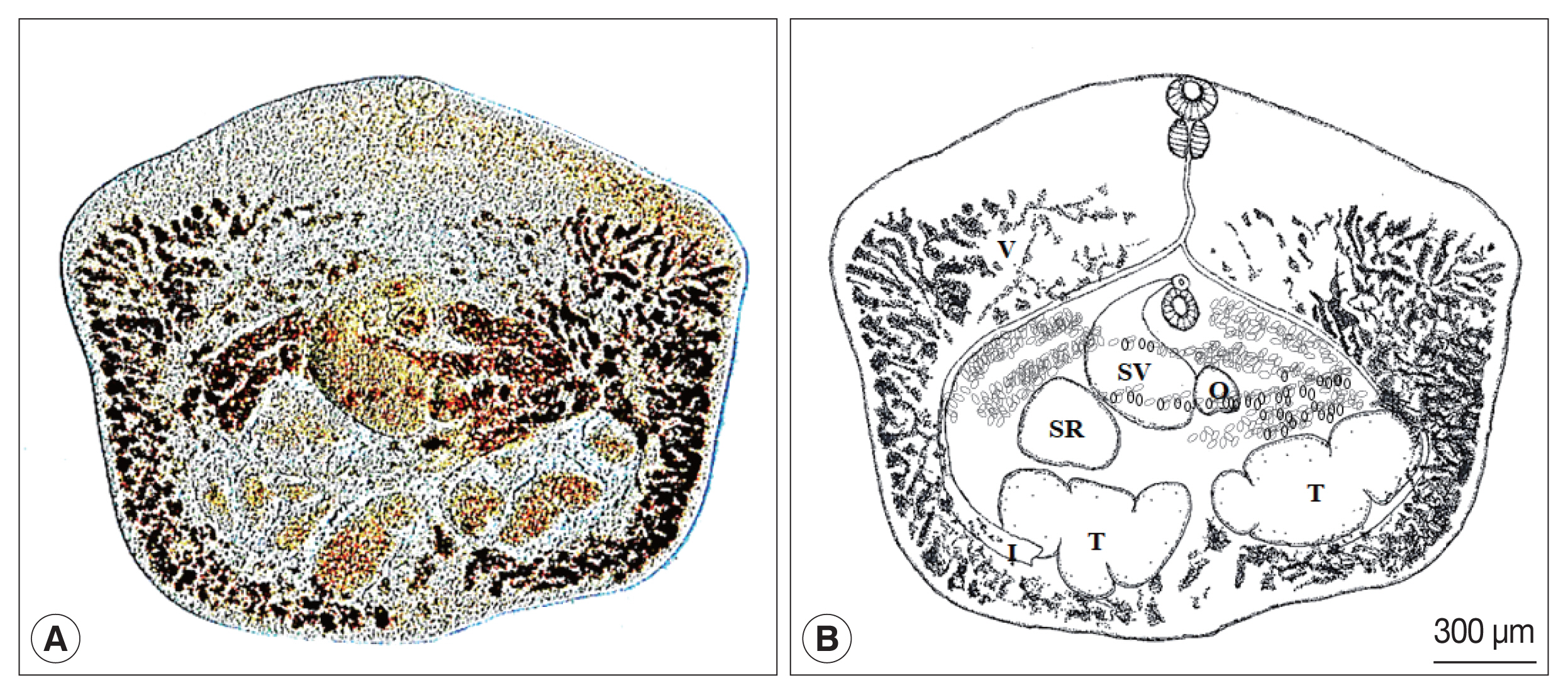
The morphological characters of heterophyid flukes in this study were as follow (Fig. 1). Body covered with many minute spines, rectangular, broader than length, 807–1,103 (1,053)×1,270–1,550 (1,460) μm. Oral sucker 91–143×100–141 μm, lying at the anterior end of the body. Pharynx nearly spherical, 79–95 (91)×85–106 (98.5) μm, and esophagus well-developed, sigmoid and long. Esophagus bifurcates at the anterior to acetabulum, and ceca follow the contour of the body to the posterior. Acetabulum 103–124 (118)×127–150 (140) μm, lying in the middle of the body. Genital atrium opens immediately anterior to acetabulum and is overhung by genital papillae. Testes symmetric or slightly diagonal, globular and lie on either side of the median at the posterior of the body. Testes 511–582 (557)×268–300 (294) μm and 468–547 (515)×254–300 (287) μm and deeply lobed. Ovary 125–141 (139)×97–124 (117) μm, heart-shaped or triangular in shape and lies slightly to the left side, anterior to left testis. Seminal receptacle 502–582 (552)×197–229 (217) μm, heart or club-shaped and lies between the right testis and seminal vesicle. Seminal vesicle 380–424 (413)×311–371 (352) μm, club-shaped, bends medially beneath the right edge of acetabulum, and opens via a short ejaculatory duct into genital pore. Genital pore and atrium located medially in front of acetabulum and covered by gonotyle. Uterus consists of 3 or 4 loops at the right side of the body and opens into genital atrium. Vitelline follicles dendritic and extend from the intestinal bifurcation to the posterior part of the body, following the ceca, but do not exceed the extra-cecal margin. Eggs 33–35 (34.5)×15–16 (15.6) μm, polar thickening and operculated.
The several species of carnivora act as definitive host of the genus Euryhelmis [1–9]. Although many heterophyid flukes from carnivora had been reported and described in Korea [10–21], there is no literature of E. squamula and their host in Korea. In this study, E. squamula was recorded for the first time as the natural infection in a raccoon dog from Korea.
In 1819, Rudolphi [22] incompletely described Distomum squamula, which later became the genus Eurysoma (Dujardin, 1845), and finally, Euryhelmis (Heterophyidae) in 1925 by Poche [23]. The Heterophyidae subfamily, Euryhelminthinae, is composed of wide-bodied trematodes. Baer [24] fully described E. squamula (type species) in 1931, and the taxonomic location of E. squamula was confirmed by Callot’s, publishing the adult measurements in 1946 [25].
Yamaguti [26] reported that the most identifiable features of the subfamily Euryhelminae are a wider body, short esophagus, almost symmetrical testes, and vitelline follicles located in the extra-cecal margin, lateral to the pharynx and testes. Further exploration of the species from genus Euryhelmis showed that the morphology, taxonomy, and occurrence of these trematodes were variable [27], but the superfamily Euryhelminae: Heterophyidae could be identified based on the following characteristics: a longitudinally or transversally elongated flat body, a variable esophagus length, the acetabulum is located a small distance in front of the middle of the body, the genital pore is immediately in front of the acetabulum and covered by a gonotyl (varying in size and structure), 1 or 2 testes are located at the posterior of the body, the ovary and seminal receptacles are transversally elongated and lie in front of the right testes, the vitelline follicles are in the lateral margins and reach behind the testes at the body’s posterior end to the intestine bifurcation level, or to the level of the pharynx and oral sucker at the body’s anterior end, the uterus is short with several convolutions between the gonotyl and transversal vitelline channels, and there is a Y-shaped excretory vesicle. The characteristic keys of Euryhelmis are the body rectangular or pyriform, testis lobed, vitelline follicles in both fore- and hind-body, natural definitive host raccoon and mink [1]. In this study, the wider body is rectangular, testis deeply lobed, vitelline follicles in both fore- and hind-body. The genital pore opening in front of the acetabulum is an important characteristic note of Heterophyidae [1], and this is same as this study.
The E. squamula morphological characteristics are as follows: the body is small, leaf-like, and broader than long, the excretory vesicle is Y- or T-shaped, there is 1 transitory testis or 2 spherical or lobate testes that are large and persist throughout adult life, the uterus relatively short and consists of 3 loops, principally situated at the left side of the body between the acetabulum and excretory vesicle, the vitelline follicles are numerous, primarily lateral, and extend from the posterior region of the body to the intestinal bifurcation, the genital atrium is immediately in front of the acetabulum, the intestinal crura extends to the posterior extremity of the body, the eggs are operculated, with or without slight polar thickening, and the adults are found in the intestine of mustelids [3]. The morphological appearance of heterophyid flukes shown in this study was well consistent with the morphological characteristics of E. squamula: the body is small, leaf-like, and broader than long. The uterus relatively short and consists of 3 loops, and the vitelline follicles numerous, primarily lateral, and extend from the posterior region of the body to the intestinal bifurcation. The intestinal crura extends to the posterior extremity of the body. The uterus consists of 3 or 4 loops at the right side of the body.
The comparison of morphometric features of the genus Euryhelmis species is compared in Table 1. The body shape of E. monochis, E. zelleri, E. squamula, and this fluke are broader than long, which differs from E. asiaticus, E. costaricensis, E. cotti, E. pacificus, E. pyriformis that are pyriform or elongate. E. monochis, E. squamula, and this fluke have the characteristic rectangular shape. E. mornochis, E. zelleri and E. cotti are smaller than E. squamula, and this fluke that identified from this study. Length and width of the E. squamula are 0.6–1 mm and 1.4–1.9 mm, respectively. In this study, the specimen’s body length and width are 807–1,103 μm and 1,270–1,550 μm, respectively. E. asiaticus, E. costaricensis, E. monochis, E. squamula and this fluke differ from E. pacificus, E. pyriformis, and E. zelleri, that the acetabulum is larger than the oral sucker. E. squamula resembles E. costaricensis in morphology (the acetabulum is larger than the oral sucker), but the E. costaricensis acetabulum is located anterior to the body length midpoint, and the E. squamula acetabulum is located at the body length midpoint. Similarly, E. asiaticus has a slender and straight esophagus and an acetabulum is larger than the oral sucker, but to find the relationships among E. costaricensis, E. squamula, and E. asiaticus require further investigations [27]. E. monochis and E. pyriformis differ from the other species by having 2 non-transitory testes. E. squamula and E. monorchis are also morphologically similar [4], but differ in the testes. E. monorchis has 1 transitory testis, and E. squamula has 2 persistent testes. Also, E. asiaticus has a slender and straight esophagus, but most of species, including E. squamula and this fluke, have sigmoid esophagus. The seminal vesicle and seminal receptacule of this this fluke are larger than other species. The fluke of this study is slightly larger than E. squamula of Grabda-Kazubska [7] in most of the measurements, but similar to E. squamula.
In this study, the flukes are small, leaf-like, broader than long, and rectangular in shape. Although, we could not obtain a properly stained specimen, the microscopic finding of non-stained heterophyid fluke revealed the features of internal organ. The acetabulum was smaller than the oral sucker, and the sigmoid esophagus bifurcates anterior to the acetabulum. The testes deeply lobed, and the vitelline follicles numerous, primarily lateral, and extend from the posterior region of the body to the intestinal bifurcation. The genital atrium is immediately located in front of the acetabulum, and the intestinal crura extend to the posterior extremity of the body. The results herein reveal that the fluke of this study is E. squamula.
E. squamula is a trematode parasite with a complex lifecycle, which require at least 3 hosts to complete it. The first intermediate host of E. squamula is a tiny, operculate snail, Bythinella hemphilli Pilsbry (Family Hydrobiidae), which develops lophocercous cercariae in the rediae after infection [28]. The second intermediate hosts are frogs and toads, such as Ascaphus truei, Rana aurora, Rana cascadae, Rana esculenta L., Rana pipiens, Rana temporaria, and Triturus cristatus Laurenti, as evidenced by E. squamula encysted metacercariae on the skin [28–30]. The final hosts of Euryhelmis are raccoons, weasels, badgers, and minks [24,29–40]. Moreover, globally, M. vison, M. putorius, Procyon lotor, Mustela frenata, Martes melampus, Martes nivalis, Martes americana, Martes sibirica, Meles anakuma, O. zibethicus, and S. bendirii palmeri are distributed. In Europe, E. squamula was reported from M. nivalis, M. putorius, M. frenata and Vulpes vulpes. In North America, E. squamula was reported from M. vision, M. americana, P. lotor, O. zibethicus, and S. bendirii palmeri [29–39], and in Asia from M. melampus and M. anakuma [40]. In this study, the adult worms of E. squamula were found in the intestine of N. procyonoides koreensis and now, N. procyonoides koreensis is new definitive host of E. squamula.
A limited point, i.e., no stained specimens were obtained, but our photographs of unstained heterophyid flukes taken before the fixation are closely identical with the characteristic morphologies of E. squamula.
In conclusion, based on the morphological comparison of 8 Euryhelmis species, the small heterophyid flukes recovered from the intestine of Korean raccoon dog, N. procyonoides koreensis, were identified as E. squamula in this study, and E. squamula is to be a new trematode fauna in Korea. Accordingly, we report for the first time that the Korean raccoon dog, N. procyonoides koreensis, serves as the definitive host of E. squamula, and E. squamula is distributed in Korea. However, there is no information on the first and second intermediate hosts of E. squamula in Korea. Further studies on what kind of animals act as the first and second intermediate hosts of E. squamula should be continued in Korea.