Warning: mkdir(): Permission denied in /home/virtual/lib/view_data.php on line 81
Warning: fopen(upload/ip_log/ip_log_2024-04.txt): failed to open stream: No such file or directory in /home/virtual/lib/view_data.php on line 83
Warning: fwrite() expects parameter 1 to be resource, boolean given in /home/virtual/lib/view_data.php on line 84 Growth and development of Pygidiopsis summa in rats and mice with a supplementary note on its morphological characters
Growth and development of Pygidiopsis summa in rats and mice with a supplementary note on its morphological characters
Jong Yil Chai,Byong Seol Seo,Soon Hyung Lee and Sung Tae Hong
Department of Parasitology and Institute of Endemic Diseases, College of Medicine, Seoul National University, Seoul 110, Korea.
Abstract
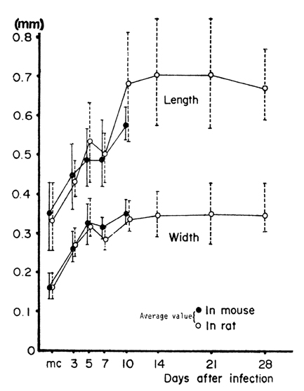
The growth and development of Pygidiopsis summa were studied in experimental rats and mice, and a special reference was given to its morphological characters differed from the type species, P. genata. The metacercariae were obtained from young mullets (Mugil sp.), and total 21 rats and mice infected each with 1,000 metacercariae. Worms of various ages of infection, from 3 to 28 days, were subjected to a microscopic observation. The worms grew rapidly and remarkably in size up to 10 days of infection, to become 0.53-0.82 mm long and 0.31-0.39 mm wide, but nearly stopped the growth thereafter. Their genital organs developed more rapidly and fully matured within 3-5 days. At 3 days several eggs were found in uterus. The presence of two groups of small spines, 5-6 on the right and 7-9 on the left side of the genital apparatus, was a new finding in this study and considered a distinct character of P. summa. The morphology of ventral sucker and intestinal ceca was also different from P. genata. This study confirms the validity of the species, P. summa.
Figures
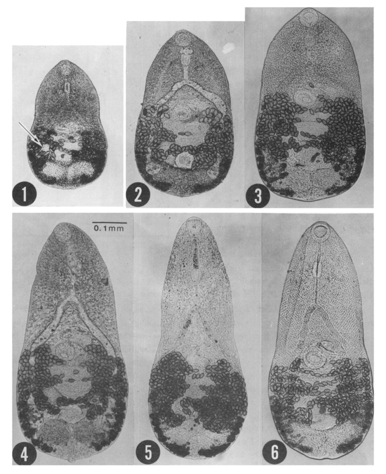
Fig. 1-6 Chronologic development of P. summa in rats and mice(unstained specimens after fixation).
1. A 3day old worm. All of genital organs are already formed and eggs are seen, but vitellaria are not fully differentiated into groups of follicles. 2. A 5 day worm. Eggs are increased in number and vitellaria begin to from groups. 3. A 7 day old worm. Eggs are greatly increased in number and vitellaria appear to have been fully differentiated. 4. A 10 day worm. Body shows its full-grown size and all organs are in their maturity. 5. A 14 day worm. Many eggs are retained. 6. A21 day worm. Note a little decreased number of eggs.
Fig. 7 Growth curves of P. summa in rats and mice (Worms older than 10 days werenot recovered from the mice).
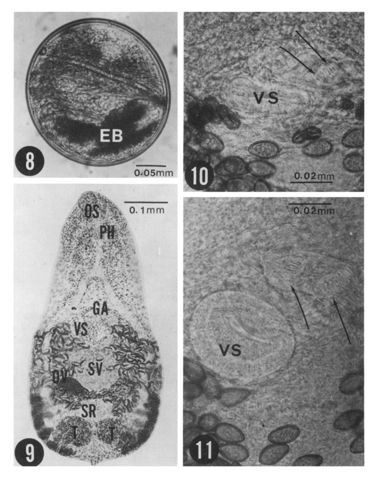
Figs. 8-11 Fig. 8. A metacercaria of P. summa collected from a mullet. The X-shaped excretory bladder(EB) is a characteristic morphology.
Fig. 9. A fully mature P. summa recovered 10 days after infection to a rat host. Acetocarmine stained specimen(OS; oral sucker, PH; pharynx, VS; ventral sucker, GA; genital apparatus, SV; seminal vesicel, and T; testes).
Fig. 10. Magnification of th middle portion of a 3 day old worm. Ventral sucker(VS) and genital apparatus or gonotyl armed with 7 minute spines on the lift side (arrow heads) and several intrauterine eggs are seen.
Fig. 11.Ibid, a 7 day worm. The gonotyls formed two groups and each armed with 5(right side) and 9(left) spines (arrow heads).
Tables
Table 1 Measurements of P. summa recovered from albino rats and mice
Table 2 Some differential characters between P. summa and P. genata
References
1.
Faust EC, et al. J Parasit 1926;13(2):91–131.
2.
Seo BS, Hong NT. Study On Metagonimus Yokogawai(Katsurada, 1912) In Korea: I. On The Metacercaria, Its Distribution In The Second Intermediate Host And The Development In The Final Host. Korean J Parasitol 1969;7(3):129–142.
3.
Ito J. Progress of Med Parasit in Japan 1964;1:317–393.
4.
Kuntz RE, Chandler AC. Studies on Egyptian trematodes with special reference to the heterophyids of mammals. I. Adult flukes, with descriptions of Phagicola longicollis n. sp., Cynodiplostomum Namrui n. sp., and a stephanoprora from cats. J Parasitol 1956;42(41):445–459.
5.
Kooss A. Centrabl f Bakt etc I Abt Originale 1907;43(5):478–490.
6.
Nishio T. Tokyo Iji Shinshi 1915;1923:1139–1142.
7.
Ochi S. Tokyo Iji Shinshi 1931;2712:346–353.
8.
Onji Y, et al. Chiba Igaku Semmon Gakko Zasshi 1916;81&82:229–249.
9.
Onji Y, et al. Chiba Igakkai Zasshi 1926;2(3):113–161.
10.
Ransom BH. Proceed US Nat Museum 1920;57:527–573.
11.
Seo BS, et al. Seoul J Med 1980;21(1):30–38.
12.
Bs Seo,et al. Seoul J Med 1981;22(2):228–235.
13.
Bs Seo,et al. Seoul J Med 1981;22(2):236–342.
14.
Witenberg G. Ann Trop Med Parasit 1929;23:131–268.
15.
Yokoagwa M, et al. Japanese J Parasit 1965;14(6):577–585.