Warning: mkdir(): Permission denied in /home/virtual/lib/view_data.php on line 81
Warning: fopen(upload/ip_log/ip_log_2024-04.txt): failed to open stream: No such file or directory in /home/virtual/lib/view_data.php on line 83
Warning: fwrite() expects parameter 1 to be resource, boolean given in /home/virtual/lib/view_data.php on line 84 Studies on intestinal tematodes in Korea XVI. Infection status of loaches with the metacercariae of Echinostoma hortense
Studies on intestinal tematodes in Korea XVI. Infection status of loaches with the metacercariae of Echinostoma hortense
Jong Yil Chai,Sung Jong Hong,Woon Mok Sohn,Soon Hyung Lee and Byong Seol Seo
Department of Parasitology and Institute of Endemic Diseases, Collge of Medicine, Seoul National University, Seoul 110, Korea.
Abstract
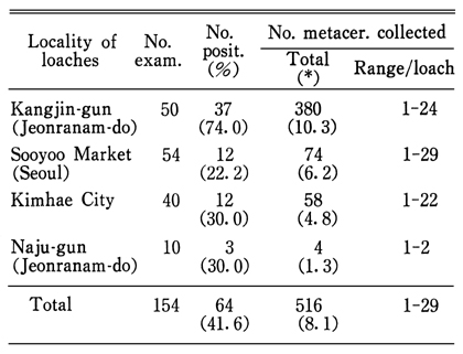
The infection status of the loaches, Misgurnus anguillicaudatus, with the metacercariae of Echinostoma hortense, was studied in Korea. A total of 154 loaches purchased at 4 local makets (Seoul, Kimhae, Naju-gun and Kangjin-gun) were examined their infection rate as well as the density and location of the metacercariae in the fish body. The results are as follews: The loaches carrying the metecercariae of E. hortense were 64 (41.6%) in total number and the metacercarial density ranged 1-29 per infected loach with an average value of 8.1. The highest infection rate and metacercarial density were obtained from the loaches purchased at Kangjin-gun, Jeonranam-do. The metacercaria of E. hortense were chiefly distributed in the distal intestinal wall and the adjacent mesentery, the perianal tissues, and the head and gill of the loaches examined. From the results, it is concluded that the loach is one of the important second intermediate hosts of E. hortense in Korea, and their infection rate and metacercarial density are considerably high.
Figures
Explanation of Figures Fig. 1. Encysted metacercaria of E. hortense, unpressed (scale: 30µm).
Fig. 2. Another metacercaria showing the characteristic structures of collar spines, suckers and excretory bladder with large corpuscles, pressed specimen (scale: 30µm)
Fig. 3. Magnification of Fig. 2. The dorsally uninterrupted collar spines (arrow heads) are seen (scale: 20µm).
Fig. 4. Another metacercaria in the perianal tissue of a loach. Note 4 end group spines (circles) at both sides of the oral sucker (scale: 20µm).
Fig. 5. An excysted metacercaria showing the oral sucker with collar spines, ventral sucker and excretory bladder. Note 4 end group spines (circles) (scale: 50µm).
Fig. 6. Magnification of Fig. 5, showing the spines of end groups (circles) and several lateral ones (scale: 20µm).
Fig. 7. Dorsal view of the oral sucker of another metacercaria showing the dorsally uninterrupted collar spines (scale: 20µm).
Tables
Table 1 Intfection status of the loaches with the metacercariae of c
Table 2 Location of E. hortense metacercariae in body of the loaches
References
1.
Arizono N, et al. Jpn J parasitol 1976;25(1):36–45.
2.
Asada S. Trans jpn Pathol Soc 1926;16:293–294.
3.
Asada S. Tokyo Iji Shinshi 1927;2527:926–930.
4.
Cho SY, Kang SY, Ryang YS. [Helminthes Infections In The Small Intestine Of Stray Dogs In Ejungbu City, Kyunggi Do, Korea]. Korean J Parasitol 1981;19(1):55–59.
5.
Kamiya H, Ishigaki K. Helminths of mustelidae in Hokkaido. Jpn J Vet Res 1972;20(4):117–128.
6.
Makino Y, et al. Jpn J Parasitol 1982;31(5):385–390.
7.
Miyamoto K, et al. Jpn J Parasitol 1983;32(4):261–269.
8.
Mori J. Tokyo Iji Shinshi 1935;No2,929:1236–1244.
9.
Okahashi K. Okayama Igakkkai Zasshi 1966;78:15–24.
10.
Ono S. Dobutsugaku Zasshi 1930;42:7–6.
11.
Park JT. Keijo J Med 1938;9(4):283–286.
12.
Ryang YS, Ahn YK, Lee KW, Kim TS, Han MH. [Two cases of natural human infection by Echinostoma hortense and its second intermediate host in Wonju area]. Korean J Parasitol 1985;23(1):33–40.
13.
Saito S, et al. Jpn J Parasitol 1982;31(4):281–287.
14.
Seo BS, et al. Seoul J Med 1980;21(1):21–29.
15.
Seo BS, Cho SY, Hong ST, Hong SJ, Lee SH. Studies On Parasitic Helminths Of Korea 5.Survey On Intestinal Trematodes Of House Rats. Korean J Parasitol 1981;19(2):131–136.
16.
Seo BS, Chun KS, Chai JY, Hong SJ, Lee SH. Studies on intestinal trematodes in Korea: XVII. Development of egg lying capacity of Echinostoma hortense in albino rats and human experimental infection. Korean J Parasitol 1985;23(1):24–32.
17.
Seo BS, Hong ST, Chai JY, Lee SH. Studies On Intestinal Trematodes In Korea: VIII. A Human Case Of Echinostoma Hortense Infection. Korean J Parasitol 1983;21(2):219–223.
18.
Seo BS, Rim HJ, Lee CW. Studies on the parasitic helmiths of Korea: I. Trematodes of rodents. Korean J Parasitol 1964;2(1):20–26.
19.
Tani S. Jpn J Parasitol 1976;24(4):262–273.
20.
Tani S. Jpn J Parasitol 1976;25(6):461–467.
21.
Tani S. Jpn J Parasitol 1979;28(1):57–62.
22.
Tani S, et al. Jpn J Parasitol 1974;23(6):404–408.