Warning: mkdir(): Permission denied in /home/virtual/lib/view_data.php on line 81
Warning: fopen(upload/ip_log/ip_log_2024-04.txt): failed to open stream: No such file or directory in /home/virtual/lib/view_data.php on line 83
Warning: fwrite() expects parameter 1 to be resource, boolean given in /home/virtual/lib/view_data.php on line 84 Electron microscopical and histochemical studies on the epicuticle of Echinorhynchus gadi (Acanthocephala)
Electron microscopical and histochemical studies on the epicuticle of Echinorhynchus gadi (Acanthocephala)
Byung Chai Cho
Department of Parasitology, School of Medicine, Kyung Hee University, Korea.
Abstract
For the purpose of observing ultrastructure of the epicuticle of Echinorhynchus gadi, the present electron microscopical studies had been made. Also the histochemical methods of Morwy, Bauer, Smith, Lison, Taft, and those of lead and uranyl acetate had been used in order to see the distribution of glycogen, mucopolysaccharides, lipid and nucleic acid in the cuticle of Echinorhynchus gadi. The results obtained by the above studies were as follows:
1. Glycogen, mucopolysaccharides were found in the outermost, middle and inner layers, especially abundant in the middle layer of the cuticle.
2. Lipid was found in the middle and inner layer, and it was found abundantly around the lacunal canal in the cuticle.
3. Nucleic acid was found around the lacunal canal in the middle layer, and also distributed in the cell nucleus of inner layer in the cuticle.
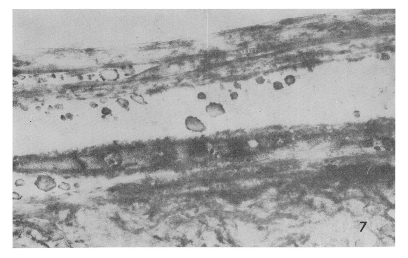
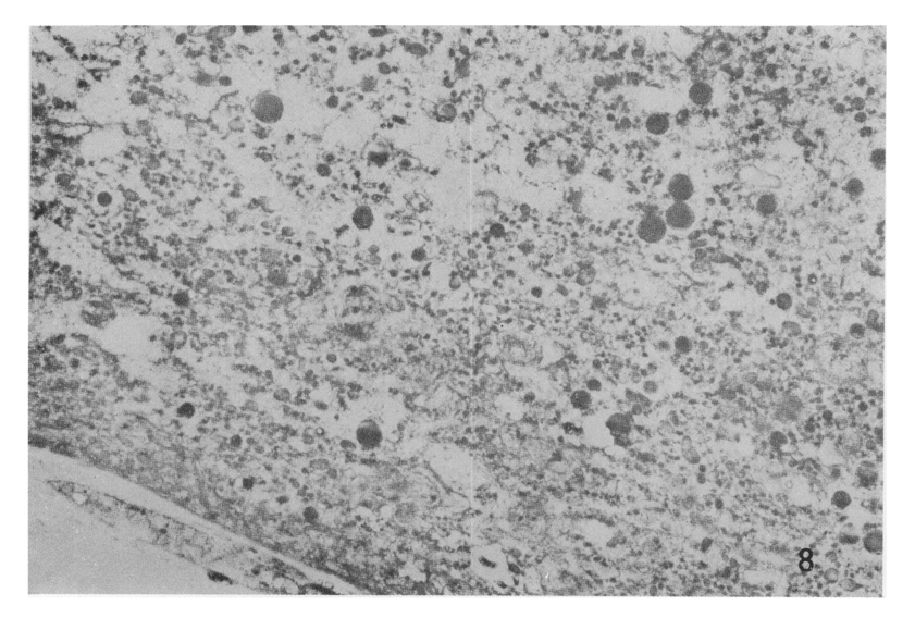
4. Electron microscopically, the cuticle of Echinorhynchus gadi had three outer layers, being outermost, middle and inner ones. The outermost layer was medium electron dense, composed with plasmalemma and filaments. The middle layer was homogeneous one which was electron pale. The inner layer, which was electron dense, consisted of felt layer and radial layer. The electron dense glycogen, lipid granules were distributed in radial layer.
Figures
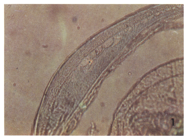
Fig. 1 Integument showed outer, middle and inner layers, which were stained with light blue. Every section had been with alcian blue. ×450
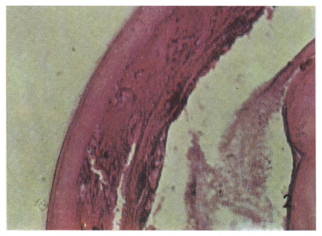
Fig. 2 Integument showed red purple, when it was stained with P.A.S. ×450
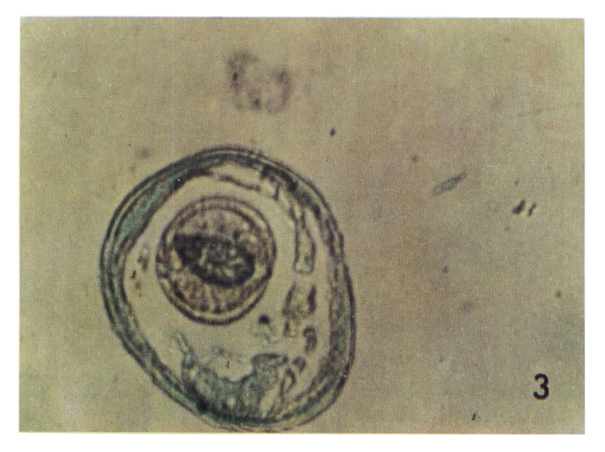
Figs. 3 Integument showed light blue and parenchymal tissue show pink, when they were stained with nile blue. ×40
Fig. 4 Outermost layer showed light blue, and middle and inner layers showed reddish pink, when they were stained with sudan black B. ×450
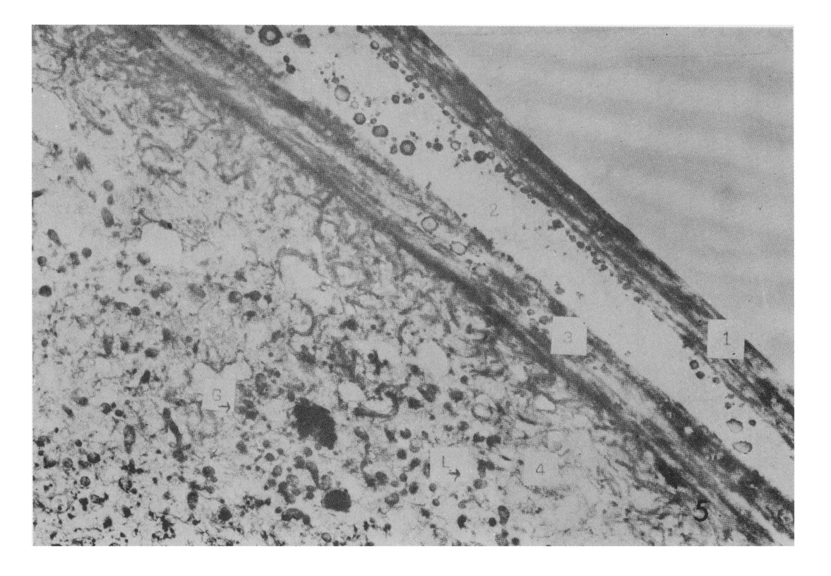
Fig. 5 Epicuticle showed three layers: (1) Outermost layer was electron dense; (2) Middle layer showed electron pale with electron dense granules; (3) Inner layer showed electron dense with plasmalemma and filaments. ×6,250