Warning: mkdir(): Permission denied in /home/virtual/lib/view_data.php on line 81
Warning: fopen(upload/ip_log/ip_log_2024-04.txt): failed to open stream: No such file or directory in /home/virtual/lib/view_data.php on line 83
Warning: fwrite() expects parameter 1 to be resource, boolean given in /home/virtual/lib/view_data.php on line 84 A study on the fine structure of Clonorchis sinensis, a liver fluke III. The prostate gland
A study on the fine structure of Clonorchis sinensis, a liver fluke III. The prostate gland
Kye Heon Jeong,Han Jong Rim and Chang Whan Kim
Department of Biology, Soon Chun Hyang College, Korea.
Department of Parasitology and Institute for Tropical Endemic Diseases, College of Medicine, Korea University, Korea.
Department of Biology, College of Science, Korea University, Korea.
Abstract
A study on the ultrastructures of the prostate gland of Clonorchis sinensis was conducted. The presence of the prostate gland in this fluke has not been known up to present time. Authors observed the ejaculatory duct epithelium including its surrounding parenchyma and found the prostate gland with the help of the electron microscope. The prostate gland was consisted of numerous unicellular glands grouped around the ejaculatory duct. The individual cell was lobulated, tapering in the direction of the ejaculatory duct. The secreting ducts of the gland penetrated into the ejaculatory duct through the muscular layer and the basement membrane, and finally opened to the lumen of the ejaculatory duct. The secreting duct had single layered microtubules along the inner wall of the duct. The secretory bodies produced by the prostate gland seemed to be moved to the lumen of the ejaculatory duct through the secreting duct. The prostate gland of this fluke was less developed than that of Fasciola hepatica but the basic structures were quite similar. There were well-developed lamellae in the epithelia of all ducts concerning passage of spermatozoa from the testes to the male genital opening.
Figures
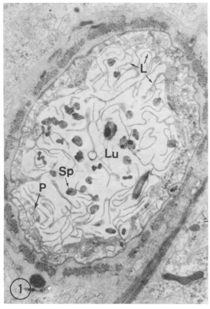
Fig. 1 Cross view of vasa efferentia. The epithelial cells have lamellae(L) at apical part of them. The protoplasmic membrane(P) is observed between the cells. There is no termination of the prostate gland. Lu, Lumen; L, Lamellae; Sp, Sperms. ×10,300.
Fig. 2 Prostate gland. The lobulated gland cells(PG) are grouped around the ejaculatory duct(EjD) and they are tapering in the direction of the duct. S, Secretory body. ×6,800.
Fig. 3 Epithelium of the ejaculatory duct. This figure shows the full and collapsed terminations of the prostate glands and the apical extension of the duct epithelium. Lu, Lumen of the ejaculatory duct; S, Secretory bodies; CM, Circular muscles; Sp, Sperms. ×7,000.
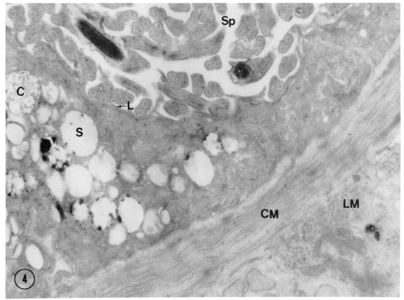
Fig. 4 High magnification of the ejaculatory duct epithelium. This shows many termination of the prostate gland with condensing vacuoles(C) supposed to be immature secretory vesicles and mature secretory bodies(S), L, Lamella; CM, Circular muscle; LM, Longitudinal muscle; Sp, Sperms. ×23,700.
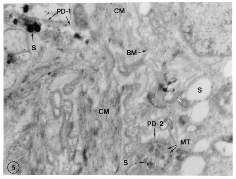
Fig. 5 Duct of the prostate gland. A secreting duct(PD-1) is penetrating into the ejaculatory duct through the muscular layer(CM) and the basement membrane(BM). The crossly sectioned duct found in the ejaculatory duct epithelium(PD-2) shows single layered microtubules along the inner wall of the duct. Certain secretory bodies(S) with high electron density are observed in the ducts. ×23,700.
References
1.
Bogitish BJ. J Parasitol 1970;56:1084–1094.
2.
Thorsell W, Björkman N. On the fine structure of the Mehlis gland cells in the liver fluke, Fasciola hepatica L. Z Parasitenkd 1965;26(1):63–70.
3.
Threadgold LT, Irwin SW. Electron microscope studies of Fasciola hepatica. IX. The fine structure of Mehlis' gland. Z Parasitenkd 1970;35(1):16–30.
4.
Threadgold LT. Electron microscope studies of Fasciola hepatica III. Fine structure of the prostate gland. Exp Parasitol 1975;37(1):117–124.
5.
Threadgold LT. Fasciola hepatica: the ultrastructure of the epithelium of the seminal vesicle, the ejaculatory duct and the cirrus. Parasitology 1975;71(3):437–443.