Warning: mkdir(): Permission denied in /home/virtual/lib/view_data.php on line 81
Warning: fopen(upload/ip_log/ip_log_2024-04.txt): failed to open stream: No such file or directory in /home/virtual/lib/view_data.php on line 83
Warning: fwrite() expects parameter 1 to be resource, boolean given in /home/virtual/lib/view_data.php on line 84 Histopathologic study on human sparganosis
Department of Pathology, College of Medicine, Seoul National University, Korea.
**Department of Parasitology, College of Medicine, Chung-Ang University, Korea.
Abstract
Based on 16 cases of human sparganosis, a histopathological study was made. There was a striking similarity among histological features of sparganosis involving different tissues. The histological change of the affected tissues was characterized by a necrotizing and granulomatous inflammation with or without worm parasite in the lesions. There was also a remarkable polymorphonuclear leukocytic mobilization, predominantly of eosinophils, plasma cells and lymphocytes in and near the lesions. Tunnel formation lined by palisading histiocytes was another charateristic feature of the host tissue reaction. These findings were quite distinguishable from those of cysticercosis which were more localized and self-limited. Several features that were prominent in section slides of sparganum worm parasite were also noted. Laminated calcospherules found in the cytoplasm of the proliferating macrophages and giant cells were of diagnostic value of sparganosis in the absence of the worm, particularly when these were accompanied with tunnel-like lesion in the host tissue.
Figures
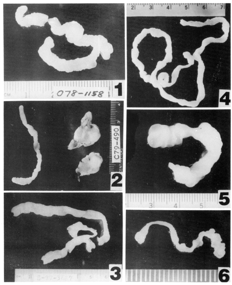
Figs. 1-6 Fig. 1. Gross appearance of the worm Sparganum in Case 12.
Fig. 2.Sparganum worm and fat tissue around the worm in Case 15.
Fig. 3.Sparganum worm removed from chest wall in Case 16.
Fig. 4.Sparganum worm in Case 14.
Fig. 5.Sparganum worm in Case 10.
Fig. 6. Anterior portion of Sparganum in Case 11.
Figs. 7-12 Fig. 7. Longitudinal section of the worm with characteristic muscle fibers.
Fig. 8. Oblique section of Sparganum, showing prominent muscle bundles.
Fig. 9.Sparganum worm showing excretory canals.
Fig. 10. Characteristic cuticular structure of Sparganum, with underlying stout muscle bundles.
Fig. 11. Cross-sectioned Sparganum and surrounding fat tissue with heavy inflammation.
Fig. 12. Higher magnification of the Sparganum worm, showing excretory system and calcospherules.
Figs. 13-18 Fig. 13. Sparganosis. Irregular cavitary necrotic lesion seen in fibrofatty tissue. Note also reaction zones around the cavity. H&E ×40
Fig. 14. Another case of sparganosis in subcutaneous tissue, showing irregular scalloping margin with active necrosis. No worm is see. H&E ×40
Fig. 15. A small focus of granuloma with degenerated Sparganum in the middle. H&E ×40
Fig. 16. Palisading histiocytic proliferation is seen around the necrotic worm. H&E ×100
Fig. 17. Higher magnification of multinucleated giant cells engulfing characteristic calcospherules of Sparganum in their cytoplasms. H&E ×360
Fig. 18. A portion of Sparganum is seen, with adjacent host tissue showing acute inflammatory cell infiltration and fibrin exudation. H&E ×100
Tables
Table 1 Age, sex and pertinent clinical findings of human sparganosis
References
1.
Chi HS, Chi JG. A Histopathological Study On Human Cysticercosis. Korean J Parasitol 1978;16(2):123–133.
2.
Cho SY, Bae JH, Seo BS. Some Aspects Of Human Sparganosis In Korea. Korean J Parasitol 1975;13(1):60–77.
3.
Mueller JF. Am J Trop Med 1938;18:303–328.
4.
Weinstein PP, Krawczyk HJ, Peers JH. Sparganosis in Korea. Am J Trop Med Hyg 1954;3(1):112–129.