Warning: mkdir(): Permission denied in /home/virtual/lib/view_data.php on line 81

Warning: fopen(upload/ip_log/ip_log_2024-04.txt): failed to open stream: No such file or directory in /home/virtual/lib/view_data.php on line 83

Warning: fwrite() expects parameter 1 to be resource, boolean given in /home/virtual/lib/view_data.php on line 84
A morphological study on spermatogenesis in the liver fluke, Clonorchis sinensis
| Home | E-Submission | Sitemap | Contact us |  
top_img
Korean J Parasito Search

CLOSE

Korean J Parasito > Volume 14(2):1976 > Article

Original Article
Korean J Parasitol. 1976 Dec;14(2):123-132. English.
Published online Mar 20, 1994.  http://dx.doi.org/10.3347/kjp.1976.14.2.123
Copyright © 1976 by The Korean Society for Parasitology
A morphological study on spermatogenesis in the liver fluke, Clonorchis sinensis
Kye-Heon Jeong,Han-Jong Rim,He-Young Yang,Woo-Kap Kim and Chang-Whan Kim
Department of Parasitology and Institute for Tropical Endemic Diseases, College of Medicine, Korea University, Korea.
Department of Biology, Korea University, Korea.
Abstract

Spermatogenesis in liver flukes, C. sinensis, was investigated by using light and electron microscopes.

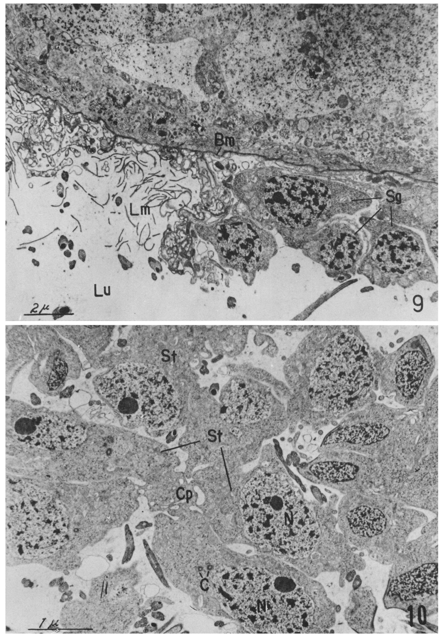
The epithelium of the testis was composed of a basement membrane, numerous lamellae protuded from the membrance and large number of spermatogonia supported by the lamellae.

The lumen of the testis was filled with numerous 8, 16 and 32-cell groups representing primary spermatocytes, secondary spermatocytes and spermatids respectively. None of cell groups with over 32 or under 8 cells was noticed.

The process of spermatogenesis is presumably as follows;

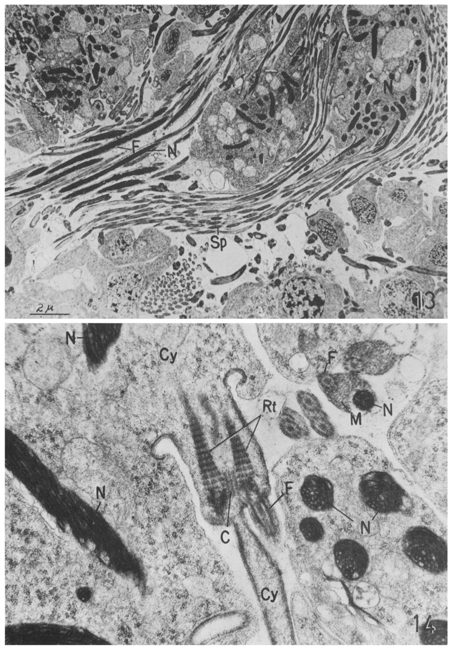
A cell group of 8 spermatogonia, attached together by a cytophore, is separated from the testis epithelium during the growth period, thus becoming primary spermatocytes. The primary spermatocytes divide to form a cell group of 16 secondary spermatocytes giving rise to a cell group of 32 spermatids through meiotic germ cell division. The spermatids begin to undergo a spermiogenesis.

The newly formed sperms remain attached together in the lumen for a while before migrating through the vasa efferentia.

Figures


Figs. 1-8
Light micrographs of germ cells,

1. Eight primary spermatocytes centrally connected by a cytophore.

2. Primary spermatocytes undergoing a meiosis.

3. Secondary spermatocytes composed of 16 cells.

4. Spermatids composed of 32 cells.

5. Spermatids undergoing a spermiogenesis. Each cell protrudes a tail(F).

6. Spermatids with elongated nuclei(N).

7. Late spermatids with elongated and bent nuclei.

8. Mature spermatozoa from seminal receptacle.


Figs. 9-10
Electron micrographs.

9. Testis epithelium composed of basement membrane(Bm), lamella(Lm), and spermatogonia(Sg).

10. Several secondary spermatocytes(St) containing centrioles (C) are connected by a cytophore(Cp).


Figs. 11-12
Electron micrographs.

11. Spermatids undergoing a spermiogenesis are still connected each other.

12. Lumen of the testis including a Sertoli cell(Sc), several spermatcytes(St) and numerous differentiation spermatozoa(Sp) in various stages.


Figs. 13-14
Electron micrographs.

13. Numerous cell groups undergoing a spermiogenesis.

14. A spermatid has a tail including two sets of filaments (F) and two rootlets(Rt).


Abbreviations
Bmbasement membrane
Ccentriole
Cpcytophore
Cycytoplasm
Ffilament
GGolgi complex
Hsperm head
Lmlamella
Lulumen of testis
Mmitochondria
Nnucleus
Rtrootlet
ScSertoli cell
Sdspermatid
Sgspermatogonia
Spspermatozoa
Stspermatocyte
References
1. Anderson WA, Weissman A, Ellis RA. Cytodifferentiation during spermiogenesis in Lumbricus terrestris. J Cell Biol 1967;32(1):11–26.
  
2. Burton PR. Fine structure of the reproductive system of a frog lung fluke. 3. The spermatozoon and its differentiation. J Parasitol 1972;58(1):68–83.
  
3. Chen PD. Trans Amer Micr Soc 1937;56:208.
 
4. Dhingra OP. Res Bull Panjab Univ 1954;44:21.
5. Dhingra OP. Res Bull Panjab Univ 1955;64:1.
6. Dhingra OP. Res Bull Panjab Univ 1955;66:19.
7. Gresson RAR. J Parasitol 1958;48:293–302.
 
8. Gresson RA, Perry MM. Electron microscope studies of spermateleosis in Fasciola hepatica L. Exp Cell Res 1961;22:1–8.
  
9. Gresson RA. Spermatogenesis of a cestode. Nature 1962;194:397–398.
  
10. Hershenov BR, Tulloch GS, Johnson AD. The fine structure of trematode sperm-tails. Trans Am Microsc Soc 1966;85(3):480–483.
  
11. Kusaura T. J Okayama Med Assoc 1966;78:929–942.
12. Lumsden RD. Microtubules in the peripheral cytoplasm of cestode spermatozoa. J Parasitol 1965;51(6):929–931.
  
13. McLaren DJ. The structure and development of the spermatozoon of Dipetalonema viteae (Nematoda: Filarioidea). Parasitology 1973;66(3):447–463.
  
14. Morseth DJ. Spermtail finestructure of Echinococcus granulosus and Dicrocoelium dendriticum. Exp Parasitol 1969;24(1):47–53.
  
15. Paik KK, et al. Kor J Electr Micr 1969;1(1):35–42.
16. Rees G. Parasitology 1939;31:417.
 
17. Rosario B. An Electron Microscope Study of Spermatogenesis in Cestodes. J Ultrastruct Res 1964;11:412–427.
  
18. Salensky W. Z Wiss Zool 1874;24:291.
19. Sato M, et al. Z Zellforsch 1967;77:232–243.
  
20. Shapiro JE, et al. J Biophys Biochem Cytol 1961;9:211–217.
  
21. Silveira M, et al. Protoplasm 1964;59:240–265.
 
22. Swiderski Z. Zool Polon 1968;18:475–486.
23. Tulloch GS, Hershenov BR. Fine structure of platyhelminth sperm tails. Nature 1967;213(5073):299–300.
  
24. von Bonsdorff CH, et al. Z Zellforsch Mikroskop Anat 1965;66:634–648.
Editorial Office
Department of Molecular Parasitology, Samsung Medical Center, School of Medicine, Sungkyunkwan University,
2066 Seobu-ro, Jangan-gu, Suwon 16419, Gyeonggi-do, Korea.
Tel: +82-31-299-6251   FAX: +82-1-299-6269   E-mail: kjp.editor@gmail.com
About |  Browse Articles |  Current Issue |  For Authors and Reviewers
Copyright © 2024 by The Korean Society for Parasitology and Tropical Medicine.     Developed in M2PI