INTRODUCTION
Human paragonimiasis has been continuously occurring in Lao People's Democratic Republic (Lao PDR) since the first case report in 1946 [1-8]. Carre (1970) analyzed the clinical manifestations in 35 Laotian paragonimiasis cases [9]. Miyazaki and Fontan (1970) reported 1 case infected by Paragonimus heterotremus adults which detected in the lung at autopsy of a Laotian man [3]. Tran et al. [7] found a cluster of paragonimiasis in a single village near Vientiane Municipality during a field survey for tuberculosis. Odermatt et al. [8] also reported 15 paragonimiasis cases among 118 residents with chronic cough in 3 villages of Hinheub District, Vientiane Province [8].
On the other hand, only 2 epidemiological studies were performed to reveal the etiological agents of paragonimiasis in Lao PDR [6,8]. Soh [6] examined 754 freshwater crabs, 524 Parathelphusa dugasti, 168 Parathelphusa sp., 62 Potamon smithianus, collected from 7 localities of Lao PDR; however, he did not find any Paragonimus metacercariae [6]. Odermatt et al. [8] detected 4 Paragonimus species metacercariae, P. harinasutai, P. bangkokensis, P. heterotremus and P. westermani, from 2 species of freshwater crabs, Potamon lipkei and Chulathelphusa brandti, collected in Nam Set River, Hinheub district, Vientiane Province [8].
As second intermediate hosts of P. harinasutai, several species of freshwater crabs, Siamthelphusa paviei [10], Larnaudia beusekomae, Larnaudia larnaudii [11], and Potamiscus smithianus [12] were reported in Thailand, and P. lipkei and C. brandti, in Lao PDR [8]. In the present study, we found P. harinasutai metacercariae in freshwater crabs, Indochinamon ou, collected from Namback District, Luang Prabang Province, Lao PDR, and adult flukes were recovered from experimentally infected dogs. We report here that I. ou is a new second intermediate host of P. harinasutai.
MATERIALS AND METHODS
Freshwater crabs (rock crabs) were collected in 8 small streams in Namback District, Luang Prabang Province, Lao PDR during 2003-2006. The collected crabs were transferred to an on-spot laboratory in Luang Prabang Province and to the laboratory in the Department of Parasitology, Hanyang University College of Medicine. The carapace of each crab was carefully separated, and body and legs were chopped with scissors and then ground in a mortar. Each ground crab was washed with tap water in a beaker. After about 15 min the supernatant was discarded. The sediment was washed with tap water or 0.85% saline until the supernatant is clear, and then examined for metacercariae under a stereomicroscope. Four specimens of crabs (2 males and 2 females) were sent to the Department of Zoology, National Museum of Nature and Science, Tokyo, Japan for species identification.
The metacercariae collected were used for morphological observations and measurements, and for experimental infection to obtain adult worms. Six dogs were each fed with 25-50 metacercariae. After 4 months, 8 Paragonimus adults were isolated from the sacrificed dog lungs. All procedures of the animal experiment followed the guidelines for the animal experiment of Hanyang University. Several adults recovered were fixed with 10% formalin under a cover glass pressure, stained with Semichon's acetocarmine and morphologically observed under a light microscope with a photo system and a micrometer. Some of them were prepared for scanning electron microscopy (SEM).
RESULTS
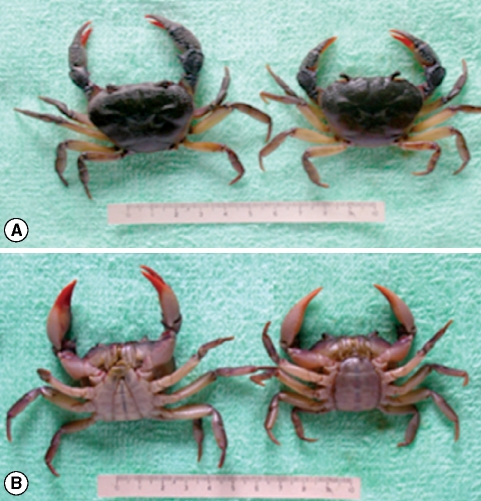
The collected crabs were 43.2 by 33.5 mm (in 2 males) and 42.3 by 32.8 mm (in 2 females) in average size. Their morphological characteristics of the carapace, the projecting anterior margin of the third maxilliped merus, and the conical and gently tapering distal segment of the first gonopod (G1) were well identical with those of I. ou (Fig. 1).
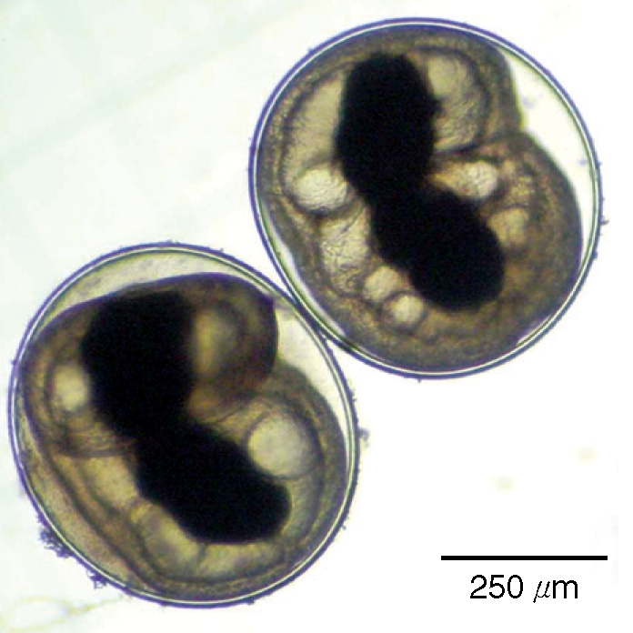
The metacercariae were round or slightly elliptical, 0.56-0.75 × 0.53-0.70 (0.67 × 0.63 mm in average) mm in size, and had a thin cyst wall of about 20 µm in thickness. They had a black excretory bladder, convoluted ceca, and some pinkish materials within the body (Fig. 2).
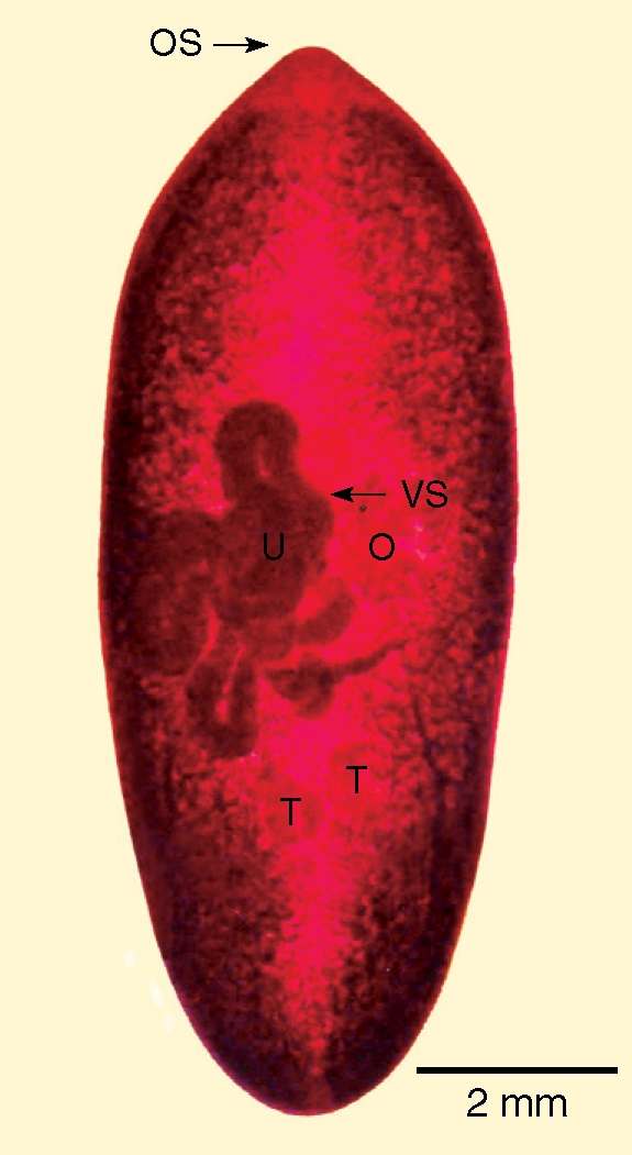
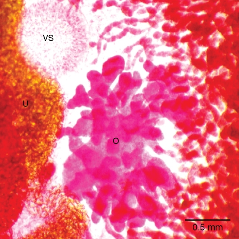
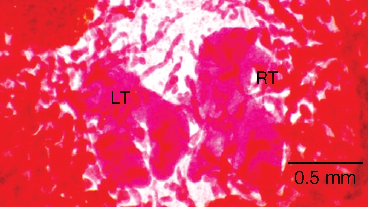
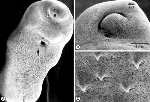
Adults were somewhat elongated, 95.2 × 36.5 mm in average size and covered with single-pointed tegumental spines (Fig. 3). The oral sucker, about 0.289 mm in diameter, was smaller than the ventral sucker, about 0.413 mm in diameter. The ovary was about 1.29 × 0.87 mm in outline, located on the right side, somewhat posterior to the ventral sucker, and moderately branched (Fig. 4). Each of the 2 testes was smaller than the ovary, and showed 5-6 lobulations (Fig. 5). Eggs were ovoid and bilaterally symmetrical in shape, 79 × 45 µm in average size, with an operculum and shoulder rims, and their shells were mostly uniform in thickness (Fig. 6). In SEM observations, P. harinasutai adults were somewhat stout and ventrally concave. The ventral sucker was larger than the oral sucker, and located at the anterior 1/3 of the body (Fig. 7A). Single-tipped tegumental spines were mainly distributed on the dorsal surface and ventro-anterior tegument between the 2 suckers (Fig. 7B,C).
DISCUSSION
In the present study, I. ou (Crustacea: Potamidae) is newly recorded as a second intermediate host of P. harinasutai. Until now, several species of freshwater crabs, P. smithianus, L. beusekomae, L. larnaudii and S. paviei were reported as the second intermediate hosts of this lung fluke in Thailand [10-12], and P. lipkei and C. brandti, in Lao PDR [8].
I. ou was described from Nam Ou at confluence with Huay Nam, 21°4'10"N 102°31"44"E, 3 km ESE of Muang Khoa, Phongsali Province, northern Laos, based only on a holotype, male specimen, and subsequently transferred to the genus Indochinamon by Yeo and Ng in 2007 [13,14]. The present specimens from Namback District, Luang Prabang Province, Lao PDR were identical with the original description in the naked carapace such as the projecting anterior margin of the third maxilliped merus, and the conical and gently tapering distal segment of the first gonopod (G1), except the shape of the second gonopod (G2). G2 of the present specimens is hooked well on the tip, whereas that of the holotype is only curved [14]. However, this morphological discrepancy may be caused by the body size. The holotype specimen is smaller and rather young, so the tip of G2 may not be hooked. In addition, the locality of the present specimens is not far from the type locality.
Four species of Paragonimus metacercariae, P. harinasutai, P. bangkokensis, P. heterotremus and P. westermani, were detected from 2 species of freshwater crabs by Odermatt et al. [8] in Lao PDR. They collected Paragonimus metacercariae in 59% of P. lipkei and 30% of C brandti examined. In a detailed examination of 7 P. lipkei crabs, 578 (84.9%) of 681 metacercariae were found to be P. harinasutai, 99 (14.5%) be P. bangkokensis, and 4 (0.6%) be P. eterotremus [8]. According to Odermatt et al. [8], and present study P. harinasutai is the dominant species in crab intermediate hosts in Lao PDR.
Paragonimiasis has been continuously occurring in Lao PDR [1-8]. As the etiologic agent of the disease, 4 Paragonimus species, P. harinasutai, P. bangkokensis, P. heterotremus and P. westermani, were proved by examination of metacercariae in the second intermediate hosts [8]. However, taxonomic studies for species identification have never been performed on the causative agent of the disease in Lao PDR except in a paper [3]. Miyazaki and Fontan [3] detected 2 flukes in the lung at autopsy of a Laotian man died of stomach cancer and identified them as P. heterotremus [3]. Accordingly, more precise and modernized methods are needed to determine the Paragonimus species causing human infections in Lao PDR.
Metacercariae of P. harinasutai were easily distinguished by the size of the cyst and the thickness of the cyst wall from those of other 3 Paragonimus species found in crab hosts in Lao PDR [8,12]. Adults recovered in the present study were identified as P. harinasutai because of the following points. (1) The oral sucker is smaller than the ventral sucker (the oral sucker of P. heterotremus is much larger than the ventral sucker). (2) Tegumental spines are single-tipped and mainly distributed on the dorsal surface and ventro-anterior tegument between 2 suckers (they are multi-pointed and distributed all over the body in P. bangkokensis, and single-pointed and distributed all over the body in P. heterotremus). (3) The ovary is moderately branched and larger than the testes (the ovary is delicately branched and smaller than the testes in P. heterotremus, and the ovary is moderately branched, almost similar with testes in size, and both genital organs very small as compared with the body size in P. bangkokensis). (4) Testes are smaller than the ovary, and 5-6 lobulated (testes are larger than the ovary and moderately branched in P. heterotremus, and testes are similar with the ovary in size and divided into 5-6 broad lobes in P. bangkokensis) [3,8,12].