Warning: mkdir(): Permission denied in /home/virtual/lib/view_data.php on line 81
Warning: fopen(upload/ip_log/ip_log_2024-04.txt): failed to open stream: No such file or directory in /home/virtual/lib/view_data.php on line 83
Warning: fwrite() expects parameter 1 to be resource, boolean given in /home/virtual/lib/view_data.php on line 84 The epidemiological studies on the filariasis in Korea I. Filariasis in Cheju-Do(Quelpart Island)
The epidemiological studies on the filariasis in Korea I. Filariasis in Cheju-Do(Quelpart Island)
Byong Seol Seo,Han Jong Rim,Soo Hyun Seong,Yong Hoon Park,Byong Chan Kim and Too Bong Lim
Department of Parasitology and Institute of Endemic Diseases, College of Medicine, Seoul National University, Korea.
Department of Internal Medicine, Cheju Provincial Hospital, Korea.
Cheju Provincial Sanitary Laboratory, Korea.
Abstract
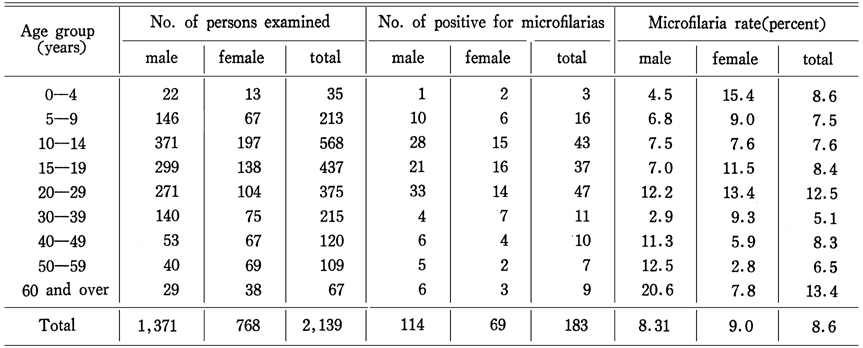
A night blood survey was carried out among inhabitants aged over 1 year from the fifteen villages throughout Cheju-Do (Quelpart Island). Blood films from 2,139 persons were examined and 183(8.6%) showed microfilariae, the incidences varying according to geographical sources are from 0.8 to 19.5 per cent. All the microfilariae found in this survey were of the nocturnal periodic Brugia malayi. The microfilarial density was 1.9 per cent of blood. The age and sex distributions of microfilaria rate in Cheju-Do were not distinctly different.
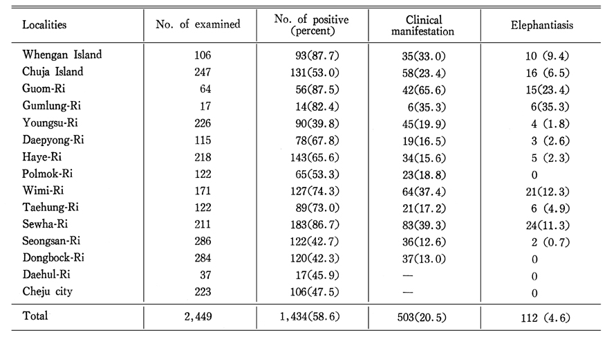
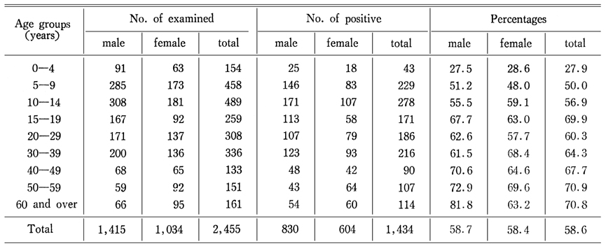
On the other hand, the intradermal test using Dirofilaria antigen (FPT antigen) and clinical survey of filariasis were also undertaken in same areas of microfilaria survey. Out of 2,449 inhabitants examined 1,434(58.6%) persons showed positive reaction of skin test, 503(20.5%) persons have clinical manifestations and 112 (4.6%) persons showed elephantiasis . It is assumed that Aedes togoi may be the most probable vector of B. malayi in the areas of Cheju-Do.
Figures
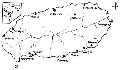
Fig. 1 Surveyed villages in Cheju-Do(Quelpart Island)
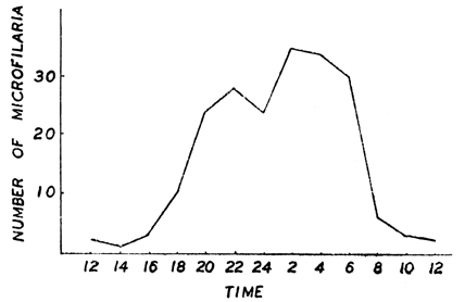
Fig. 2 Curves of microfilaraemia density (20 cmm) at various times.
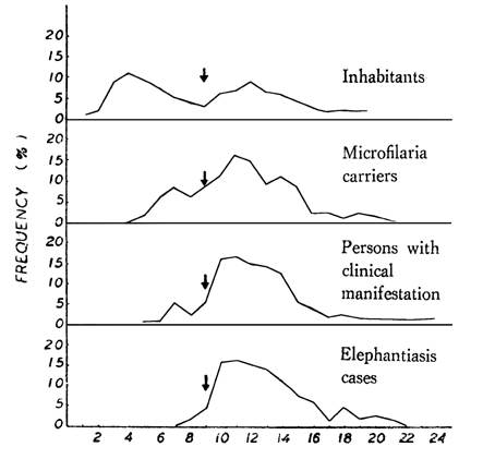
Fig. 3 Age and sex distributions of elephantiasis cases.
Fig. 4 Frequency distribution curves of skin test used FPT antigen among the inhabitants of Quelpart Island, microfilaria carrier, persons with clinical manifestation and elephantiasis cases.
Tables
Table 1 Microfilarial survey in Cheju-do(Quelpart Island)
Table 2 Age and sex distributions of microfilaria rates in Cheju-do(Quelpart Island)
Table 3 Results of intradermal test used FPT antigen of D. immitis and clinical manifestations of flariasis in Cheju-do
Table 4 Age and sex distributions of persons examined by intradermal test (EPT antigen) in Cheju-do(Quelpart Island)
References
1.
Kagan IG. A Review of Immunologic Methods for the Diagnosis of Filariasis. J Parasitol 1963;49:773–798.
2.
Lee KT. Bull Nat Inst Health Seoul Korea 1961;4(1):107–111.
3.
Senoo T, Lincicome RD. Malayan filariasis; incidence and distribution in Southern Korea. U S Armed Forces Med J 1951;2(10):1483–1489.
4.
Tada I, et al. Jap Jour Parasitol 1964;13(5):427–434.