Warning: mkdir(): Permission denied in /home/virtual/lib/view_data.php on line 81
Warning: fopen(upload/ip_log/ip_log_2024-04.txt): failed to open stream: No such file or directory in /home/virtual/lib/view_data.php on line 83
Warning: fwrite() expects parameter 1 to be resource, boolean given in /home/virtual/lib/view_data.php on line 84 The experimental studies on Capillaria hepatica
Department of Parasitology and Institute of Endemic Diseases, College of` Medicine, Seoul National University, Korea.
Abstract
Capillaria hepatica is an extremely common parasite of rats. Several human cases have also been reported from various parts of the world and recently these aroused the clinical interests.
The present study was undertaken to investigate the biological observations of C. hepatica and the changes occurring in blood picture and serum protein in the experimentally infected hosts.
The source of C. hepatica obtained from the deposit of non-embryonated eggs encapsulated in the liver of house rats(Rattus norvegicus) in Seoul. The eggs isolated from the infected liver tissues by the freshly prepared artificial gastric juice at 37℃ and embryonated in the incubator 27° to 30℃ for four to five weeks.
For the observation of migratory pathway to the liver, ten mice were infected orally with 1,000 embryonated eggs of C. hepatica, and another ten mice were infected intraperitoneally. No larvae were found in the washings of peritoneal cavity after oral infection, but after the third day of infection, the larvae were isolated from liver tissues. These indicated that the majority of larvae are transported to the liver by the hepatic portal system. On the other hand, 1,000 embryonated eggs of C. hepatica were inoculated into the peritoneal cavity of mice by mantoux syringe containing antibiotics. One third of inoculated eggs hatched out in the peritoneum during two days after inoculation, hatched in the peritoneal cavity invade directly to the surface of liver.
Twenty white rats were infected orally with 1,000 to 2,000 embryonated eggs for the study of the development of C. hepatica in the liver and histopathological changes of the infected liver in the course of infection.
C. hepatica in the liver of white rats developed rather slowly at the first tenth day after infection, but at the 13th day developed rapidly in its size. The worms were sexually differentiated at the l7th day after infection. At the 20th fully formed eggs appeared in the white or yellowish lesions on the surface of rat liver and they are also found in uterine tubule of the female worm. After the 33rd day, male worm disappeared and only female worms packed with eggs were detected in the liver tissues. However the long hair-like tightly coiled worms were also usually found in the hepatic cysts, and the degenerated or dead worms were observed in the small cysts on the surface of the liver at the 59 th day after infection.
Microscopical examination on the first week after infection revealed inflammatory reactions with the dilatation of central vein, Kupffer cell mobilization, focal necrosis and perivascular infiltration. After two weeks of infection granulomatous inflammation were observed around or adjacent to the worms in the lobules. The worms are surrounded by macrophages, multinucleated giant cells, a dense infiltration of lymphocytes, monocytes, neutrophils and, especially, eosinophils. After the third and fourth week, the microscopical findings of infected rat livers have shown proliferation of connective tissues and regeneration of liver cells. During the fifth to sixth week after infection, rat liver showed marked proliferation of fibrous connective tissues encapsulated the worms and massive deposition of the eggs. At the later time the liver reveals many pseudolobules which are caused by postnecrotic cirrhosis. These are irregularly subdivided into lobule by a fibrous septum. The worms were fragmented by the phagocytes and encapsulated by connective tissues. And then finally they appeared to be replaced by the calcium-like material. The liver shows typical cirrhosis after the eighth week after infection.
In order to investigate the changes of blood picture and serum protein components of rabbits infected with C. hepatica, twenty rabbits were divided into four groups by the doses of eggs. Group A was given doses of 1,000 embryonated eggs, group B 5,000 eggs, group C 10,000 eggs and group D 30,000 eggs.
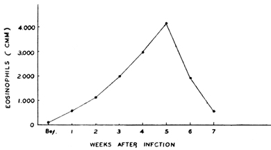
The pictures of blood especially leukocyte and eosinophil counts and of serum protein were checked every week for ten weeks in the course of infections. The marked elevation of the leukcocyte, eosinophil counts and percentage of eosinophils was observed at the sixth to the seventh week in the course of infection in all groups of rabbits. At the tenth week after infection a decrease was shown in their counts. However in the heavily infected groups (Group C & D) these values persisted relatively in high levels even thereafter. In the white rats given doses of 1,000 to 2,000 eggs, eosinophil counts increased to the peak at the fourth week and decreased at the seventh week after infection.
The changes in serum protein components of infected rabbits were investigated by paper electrophoresis. Blood collections were done by the cardiac puncture in the early morning. Serum total protein was determined by Biurets method, serum protein fractionating and A/G ratio by paper electrophoresis using Whatman No.l filter paper and barbital buffer (pH 8.6, ionic strength 0.06). Total protein increased at the sixth and seventh week after infection and the albumin and A/G ratio had decreased significantly in the heavily infected groups at the fifth and sixth week. The alpha-globulin and beta-globulin were not significant in the lightly infected groups(Group A and B), but they decreased after seventh week in the heavily infected groups. The gamma-globulin and gamma/A ratio of the heavily infected groups were significantly increased at fifth to seventh week.
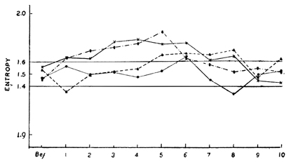
Statistically the calculation of entropy was applied to the data obtained in all groups. In the lightly infected groups, the entropy was included almost in the normal ranges, however in the heavily infected groups it was excluded from the normal range during the first to eighth week after infection.
Figures
Fig. 1 Fluctuations of white blood cells, eosinophils and percentages of eosinophils in infected rabbits.
Fig. 2 Fluctuation of eosinophils in infected rats.
Fig. 3 Variation of total protein, albumin, alpha-, beta-, gamma-globulin, A/G ratio, and γ/A ratio in infected rabbits.
Fig. 4 The curves of statistical calculated Entropy of serum protein in infected rabbits.
Fig. 5 Infected liver with Capillaria hepatica showing white or yellowish lesions of the egg deposits on the surface. (35 days after infection)
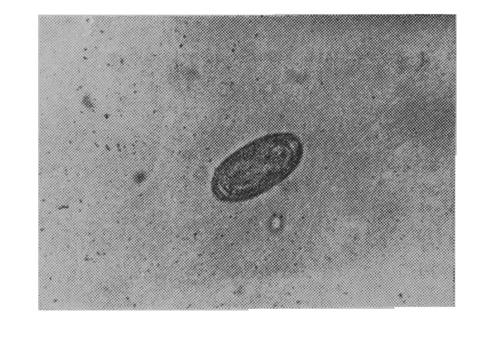
Fig. 6 Embryonated egg of C. hepatica after incubation of 45 days.
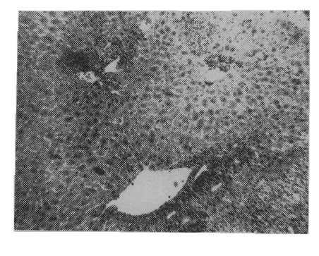
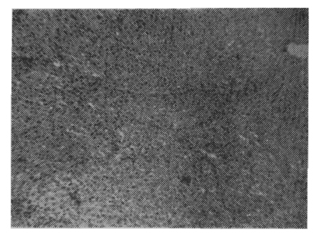
Fig. 7 The H & E stained section of the infected rat liver showing moderate infiltration of small round cells around the central veins. (7 days after infection)
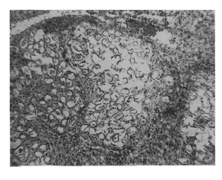
Fig. 8 The H & E stained section of the infected rat liver showing granulomatous lesion containing numerous giant cells in hepatic lobules. (17 days after infection)
Fig. 9 The H & E stained section of the infected rat liver showing massive deposition of eggs surrounded by actively proliferationg fibrous connective tissues. (42 days after infection)
Fig. 10 The H & E stained section of the infected rat liver showing postnecrotic cirrhosis consisting with irregularly subdivided lobules by fibrous septa. (7 days after infection)
Tables
Table 1 Variation of white blood cells and eosinophil level of rabbits infected with embryonated eggs of Capillaria hepatica.
Table 2 Variation of serum proteinf ractions of rabbits infected with embryonated eggs of Capillaria hepatica.
References
1.
Baba SK, et al. Jap Jour Parasit 1956;5(2):228.
2.
Bancroft TL. Jour and Proc Roy Soc New-South Wales 1893;27:86–90.
Behrer MR. Hypereosinophilia with eosinophilic granuloma of the liver associated with ascaris infestation. J Pediatr 1951 May;38(5):635–640.
7.
Brosius OT, Thomas EE, Brosius B. Capillaria hepatica a case report. Trans R Soc Trop Med Hyg 1948;42(1):95–97.
8.
Calle S. Parasitism by Capillaria hepatica. Pediatrics 1961;27:648–655.
9.
Cochrane JC, Sagorin L, Wilcocks MG. Capillaria hepatica infection in man; a syndrome of extreme eosinophilia, hepatomegaly and hyperglobulinaemia. S Afr Med J 1957;31(30):751–755.
10.
Dive GH, et al. Roy Army Med Corps 1924;43(1):1–4.
11.
van Dommelen C, Schulte MJ, Brandt K, van Leeuwen, Wadman SK. Abnormally low α2- and β-globulin levels in serious hepatic insufficiency. Acta Med Scand 1959;165:211–216.
12.
Engler G, Sanchez G. Capillaria hepatica, Bancroft, 1893; a case report. Trans R Soc Trop Med Hyg 1950;43(4):443.
13.
Evans AS, Stirewalt MA. Serologic reactions in Schistosoma mansoni infections. III. Ionographic fractionation of sera of mice with progressive disease. Exp Parasitol 1957;6(1):8–17.
14.
Ewing GM, Tilden IL. Capillaria hepatica; report of fourth case of true human infestation. J Pediatr 1956;48(3):341–348.
15.
Faust EC, et al. J Parasitol 1935;21:332–336.
17.
Franklin MH, et al. J Lab and Clin Med 1948;33:435.
18.
Fulleborn F. Archiv f Schiffs u Tropenhyg 1924;28:48–61.
23.
Hall MC. Proc US Nat Mus 1916;50:1–258.
26.
Kachi G, et al. Japan Path Soc 1924;14:176–177.
28.
Kallichurum S, Elsdon-Dew R. Capillaria in man. A case report. S Afr Med J 1961;35:860–861.
30.
Kitsuki T. Igaku Kenkyu 1958;28(5):1370–1394.
31.
Kraut N. An electrophoretic study of sera from rats artifically infected with and immunized against the larval cestode Cysticercus fasciolaris. J Parasitol 1956;42(2):109–121.
32.
Luttermoser GW. Am J Hyg 1938;27:275–289.
33.
Luttermoser GW. Am J Hyg 1938;27:321–340.
34.
MacArthur WP. Proc Roy Soc Med 1924;17(10):83–84.
35.
McQuown AL. Capillaria hepatica; report of genuine and spurious cases. Am J Trop Med Hyg 1950 Sep;30(5):761–767.
36.
McQuown AL. Capillaria hepatica. Am J Clin Pathol 1954;24(4):448–452.
37.
Martin NH. An electrophoretic study of the components of the serum proteins in cirrhosis of the liver. Br J Exp Pathol 1949;30(3):231–236.
38.
Milburn CL Jr, Ernst KF. Eosinophilia-hepatomegaly syndrome of infants and young children; report of a case due to invasion of liver by Nematode larvae. Pediatrics 1953;11(4):358–367.
40.
Momma K. Ann Trop Med Parasit 1930;24(1):109–113.
41.
Nishigori M. Jour Med Assoc Farmosa 1925;247:3–4.
43.
Piazza R, et al. Rev Inst Med Trip Sao Paulo 1963;5(1):37–41.
44.
Popper H, Bean WB, de la Huerga J, Franklin M, Tsumagari Y, Routh JI, Steigmann F. Electrophoretic serum protein in hepatobiliary disease. Gastroenterology 1951;17(2):138–162.
46.
Roberts S, White A. Studies on the origin of the serum proteins. J Biol Chem 1949;180(2):505–516.
47.
Romerogarcia FJ, et al. Bol Medi Hosp Infantile Mexico 1962;3(3):185–190.
48.
Satoskar RS, Lewis RA, Gaitonde BB. Electrophoretic studies of the plasma proteins in virus hepatitis. J Lab Clin Med 1954;44(3):349–352.
49.
Seo BS, Rim HJ, Lee CW, Yoon JS. Studies On The Parasitic Helminths Of Korea: II. Parasites Of The Rat, Rattus Norvegicus Erxl. In Seoul, With The Description Of Capillaria Hepatica (Bancroft, 1893) Travassos, (1915). Korean J Parasitol 1964;2(1):55–62.
50.
Shim BS. ROK Air Force Jour of Aviation Medicine 1954;2(2):18–27.
51.
Shorb DA. Jour Parasit 1931;17:151–154.
52.
Skrjabin KI, et al. Russian J1 Trop Med 1929;7(7):449–450.
53.
Travassos. Mem Inst Oswaldo Cruz 1915;7(2):146–172.
56.
Ward RL, et al. Bull Tullane Med Fac 1959;19:27–33.
57.
Wright WH. Am J Trop Med 1938;18:329–330.
58.
Zaiman H, Howard CJ, Drolette B. Eosinophilia in rats infected with Trichinella spiralis. Exp Parasitol 1962;12:253–262.