INTRODUCTION
Ticks have been recognized as hematophagous ectoparasites distributed worldwide. A lot of species are vectors of viral, rickettsial, bacterial, and protozoal diseases of birds and mammals, including humans [1,2]. Identification of causative tick is important in making a diagnosis in tick-related dermatoses. In the Republic of Korea, hard ticks which belong to the Family Ixodidae are responsible for most tick-related diseases. Since the first human case of tick bite was reported in 1982 [3], the total number of cases recorded in the literature reached approximate 40 [4,5]. All ticks infested in Korea belonged to the genus Ixodes or Haemaphysalis [6,7]. No Amblyomma case has yet been recorded in Korea although approximately 100 species of the genus Amblyomma were recorded worldwide. Here, we report a human case of typical infestation with an Amblyomma testudinarium (Koch, 1844) adult worm in a Korean woman.
CASE RECORD
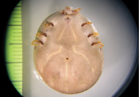
In July 2009, a 74-year-old Korean woman presented to our dermatology clinic with a large mass on her left inguinal region. She was a typical rural housewife frequently worked in the field near her house. On admission she complained of mild discomfort and itching sensation of the skin around the bite area. Laboratory examinations were normal or negative for the following tests: hemoglobin 14.1 g/dl, hematocrit 43.3%, white blood cell 8,210/mm3 (neutrophil 69%, lymphocyte 25%, monocyte 6%), erythrocyte sedimentation rate 6 mm/hr, serum glutamic pyruvic transaminase 30 IU/L, glucose 127 mg/dl. Ten days prior to presentation, the patient began to note the presence of a mass which she thought to be benign. In the clinic, we found a dark green, round, 23 mm organism whose head was partially burrowed under the skin. The organism was removed using a pair of blunt forceps. It was grasped close to the skin and steady pressure applied, pulling the tick straight out perpendicularly to the skin. When the organism was removed by plucking, its mouth part was torn. It was a grossly huge, round, and 8-legged tick (Fig. 1). To prevent inflammatory reaction due to the remnant mouth part, bite wound was surgically excised and immediately cleansed with disinfectant. The surgical wound was gradually resolved without further complications. Six months later, the patient was well and then follow-up communication was discontinued. She was probably attacked by the tick while working to collect vegetables and edible sprouts of wild grass in her vegetable garden located in the suburb of Suncheon City, Jeollanam-do, where she had been residing. She recently had not traveled to any foreign country as well as any domestic area, including Jeju Island.
DESCRIPTION OF THE TICK
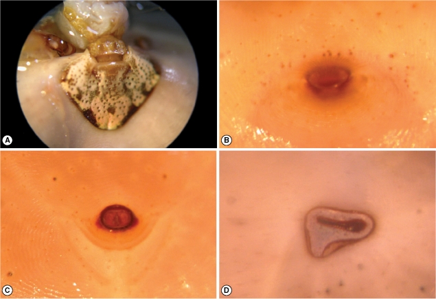
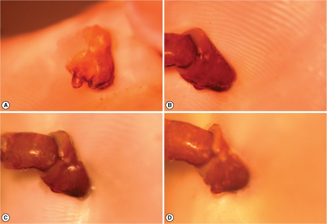
The isolated tick was fixed with formalin and then stereoscopically examined to determine its species. The body was 23 mm in length, 19 mm in width, and 12 mm in height (Fig. 1). It was widest in the region of spiracular plate. The festoons were faintly observed at the posterior margin of the body. The small ornamented scutum was seen as a small shield behind the capitulum on the back. The scutal punctations were somewhat irregular; dark brown spots on bright yellow background. The eyes were located on the lateral edges of the scutum but not projecting beyond scutal contour. Although greater part of the mouth including the hypostome was torn off, the left pedipalp only was relatively well preserved and had characteristic appearance; especially long article 2 with approximately 2.5 times as long as article 3 (Fig. 2A). A genital aperture and an anus were seen on the ventral side. The genital aperture was located at the level between coxa I and coxa II (Figs. 1, 2B). The anus was located at the level posterior to the spiracular plates and embraced by Y-shaped anal groove posteriorly (Figs. 1, 2C). The spiracular plate, comma-shaped, was located on the ventrolateral surface posterolaterally to the coxa IV (Figs. 1, 2D). The tick had 4 pairs of legs (Figs. 1, 3). The coxa I had 2 medium sized subequal spurs, external spur was slightly longer than internal spur (Fig. 3A). The coxa II had a single salient ridgelike spur which was much broader than long (Fig. 3B). The coxa III with a single spur was very similar to coxa II (Fig. 3C). The coxa IV had a single spur which was longer than those of the coxae II and III (Fig. 3D). The tarsi III and IV had 2 subapical ventral spurs. Base upon these morphological features, this tick was identified as a female adult of A. testudinarium (Table 1).
DISCUSSION
Ticks are blood-sucking arthropods that parasitize various species of vertebrates in almost every region of the world. In the Republic of Korea, about 40 human cases have been reported since 1982 [3-5]. The causative ticks reported in Korea were identified as Ixodes nipponensis, I. ovatus, I. monospinosus, I. persulcatus, Haemaphysalis flava, and H. longicornis. The remainders were Ixodes as the genus level. Among these 6 species, I. nipponensis was the most common species responsible for human bites in Korea [6,7].
Identification of ticks is mainly based on morphological characteristics of the specimen. The genus Amblyomma ticks are large and beautifully ornamented with long mouth parts; possessing eyes and festoons [8]. The large-sized tick from the present case was round in shape. Close examination under a stereomicroscope showed the capitulum of the anterior portion of the body, the scutum of the dorsal portion, and the spiracular plate, genital aperture, anus, and 4 pairs of legs of ventral portion. The pedipalp of the capitulum was slender with the second segment much longer than the third. The scutum was quite ornate in comparison to other parts of the body and had non-protruding eyes at the edge. The comma-shaped spiracular plate was located posterior to the coxa IV. Genital opening was located anterior to the level of the coxa II. The anus was surrounded posteriorly by the anal groove. The coxa of the first leg had subequal 2 spurs (the external one was slightly larger than internal). The spine of coxa on the fourth leg was longer than those on the second and third legs. The tarsi of the third and fourth legs had 2 distinct subapical ventral spurs. These characteristics were typical features of the adult female of A. testudinarium [8-11].
Ticks are usually 1 to 9 mm long before engorgement. They feed for 7 to 12 days for a full engorgement and reach 20 mm in length after feeding [1]. The genus Amblyomma is one of the largest ticks among the hard ticks. In case of A. testudinarium, the size of adult females varies from 5 to 20 mm depending on the stages of engorgement. However, this species may feed for 30 days or longer and reach bigger than 20 mm in length [9,11]. In the present case, hospital admission was quite delayed. The tick fed over additional 10 days after detected by the patient and was inordinately engorged. It measured 23 mm long, which was bigger than any other human ticks reported in Korea.
The present tick would have been in situ for at least 1 month. During that time, the patient had some itching and mild pain of the skin around the bite area, but no clinical symptoms of tick-borne infection, including Lyme disease and rickettsiosis, occurred. Ticks can also cause inflammatory problems in the host without transmitting infection [1,12]. When the head part was broken off in the removal process, more careful attention should be given to the bite wound. Therefore, small excision was performed around the biting wound in the present case. Her symptoms disappeared gradually after the removal of the tick and the surgical wound was healed in a week.
The frequent location involved by A. testudinarium is known to be the axillary, anogenital, or inguinal region, which are rich in apocrine glands [13]. In the present case, tick bite was found on the inguinal region. According to the patient's recall of daily life, she often undressed for urination while working at the vegetable garden nearby her home. It appears that the tick attached itself to her clothing or skin when she was undressed.
A. testudinarium is known to be a tropical tick and found mainly in the Indian Peninsula, South East Asia, including Myanmar, Thailand, Malaysia, Indonesia, the Philippines, Taiwan, and Japan [8]. In Japan, A. testudinarium accounts for 10% of all tick bite cases. It is especially important in southwestern areas (below about 36° North latitude), including Kinki, Shikoku, Kyushu, and Chugoku [10,14,15]. The geographical limitation of A. testudinarium in the southwestern part of Japan implies that the warm temperature with heavy rainfall and the subtropical vegetation fauna may be a proper environmental condition for this tick. In Korea, A. testudinarium has been known to be distributed in Jeju island (between 33 to 34° North latitude), which is located in the South Sea far from the Korean Peninsula [8]. However, this species was also collected in a southern coastal area of Jeollanam-do during a Haemaphysalis phasiana survey using tick drags [16]. In the present case, the patient has been living in a suburban or rural area of Suncheon City, approximately 400 km south of Seoul, Korea. It is an agricultural and industrial city nearby the sea at Suncheon Bay (35° North latitude) facing the South Sea. This area has a high temperature and humidity oceanic climate. Therefore, A. testudinarium may inhabit Suncheon City.