INTRODUCTION
Neospora caninum (Apicomplexa; Sarcocystidae) is a protozoan that mainly causes dermatological and neurological disorders for which the dog is the definitive host [1]. In cattle, the intermediate host of this parasite, N. caninum causes a range of economic losses that are associated with abortion and a decline in milk production [2]. N. caninum was first isolated in 1988 from the tissues of a naturally infected dog [3,4]. Since then, new strains have been isolated, such as NC-1 [5,6].
To elucidate the pathogenesis of infection with this parasite in the central nervous system (CNS), our group developed an in vitro model of N. caninum infection in primary glial cell cultures [7]. In this model, it was observed that astrocytes and microglia responded to infection by producing proinflammatory and regulatory cytokines, i.e., tumor necrosis factors-α (TNF-α) and IL-10, as well as the neurotoxic free radical nitric oxide (NO) [7,8,9]. These results show that glial cells are targets of N. caninum and that astrocytes may contribute to the CNS immune responses to the parasites.
Flavonoids are naturally occurring polyphenolic compounds that are present in a variety of fruits, vegetables, cereals, teas, wines, and fruit juices. They are secondary metabolites formed in plants from the aromatic amino acids phenylalanine and tyrosine as well as from malonate [10]. In Brazil and several other countries, antibacterial, antiparasitic, antifungal, and antiviral properties have been attributed to flavonoids [11,12,13]. Studies have shown that the flavonoid quercetin is able to inhibit the growth of Leishmania danovani [14], L. panamensis [15], and L. amazonensis [16]. Apigenin and the biflavone fukugetin also displayed considerable inhibition of Trypanosoma cruzi [17,18]. The isoflavone genistein efficiently blocked host cell egress of Toxoplasma gondii by more than 50% [19].
Flavonoids have been shown to modulate glial cell responses [20]. Recent findings from our group showed that flavonoid rutin induces activation of astrocytes and microglia and production of TNF-α and NO [21]. Thus, in the present study, we evaluated whether the treatment with flavonoids induces glial activation and interferes with N. caninum infection and proliferation.
MATERIALS AND METHODS
All of the experimental procedures were performed in accordance with the standards of the Ethics Committee on Animal Care of Bahia Federal University, Brazil.
Glial cell cultures
Mixed primary cultures of astrocytes and microglia were prepared according to the method of Cookson et al. [22] and modified by Silva et al. [21]. Newborn Wistar rats were decapitated on postnatal day 0-2, and their cerebral cortices were removed. After mechanical dissociation, the obtained cells were cultured in Dulbecco's modified Eagle's medium (DMEM, Cultilab, Campinas, São Paulo, Brazil) supplemented with 6.25 g/ml gentamicin, 2 mM L-glutamine, 0.011 g/L pyruvate and 10% foetal calf serum (Cultilab, Campinas, São Paulo, Brazil). The cells were seeded in 100 mm polystyrene culture plates (TPP) at a density of 3×10⁶ cells/plate. After 2-3 weeks in culture, the cells were trypsinized and replated in microtiter dishes with 40 mm, 24 or 96 wells, depending on the experiment, at a density of 1×10⁴ cells/cm2.
Neospora caninum cultivation and infection
Vero cells (from African green monkey kidneys) were maintained in DMEM enriched with 5% fetal bovine serum at 37℃ under 5% CO2, with regular changes every 48 hr. After the formation of a confluent cell monolayer, the Vero cells were infected with tachyzoites of N. caninum strain NC-1 [3,4], which was obtained from the Laboratory of Parasitology at the Veterinary Medicine Hospital of UFBA, Brazil. Glial cell primary cultures pretreated with flavonoids for 24 hr were infected with N. caninum tachyzoites according to the protocol described by Pinheiro et al. [7,8]. Briefly, at the time of infection, the Vero cells were mechanically injured using a sterile mini-rod, and the parasites were purified by exclusion chromatography in a Sephadex G25 column (Sigma, St. Louis, Missouri, USA). The number of parasites was counted, and a parasite/cell ratio of 1:1 was adopted for glial cell infection.
Flavonoid treatments
Glial cells were treated individually with 10 different flavonoids: 7-dimethoxyflavone (F1); 7,8-dihydroxyflavone (F2); 3',4'-dihydroxyflavone (F3); chrysin, 5,7-dihydroxyflavone (F4); apigenin, 4',5,7-trihydroxyflavone (F5); luteolin, 3',4',5,7-tetrahydroxyflavone (F6); kaempferol, 3,4',5,7-tetrahyroxyflavone (F7); fisetin, 5-deoxyquercetin, 3,3',4',7-tetrahydroxyflavone (F8); quercetin, 3,3',4',5,6-pentahydroxyflavone (F9); or rutin, 3 ramnoglicoside of 3,3',4',5,6-pentahydroxyflavon (F10). These compounds were dissolved in dimethylsulfoxide (DMSO) and diluted in culture medium to a final concentration of 50 µM.
To study the effect of flavonoids in uninfected conditions, glial cells were treated with each flavonoid separately for 24 or 48 hr. To investigate whether pretreatment with flavonoids interferes with NC-1 infection, another group of glial cell cultures were treated with flavonoids 24 hr before infection with N. caninum and analyzed at 24 hr post-infection, representing 48 hr of treatment with flavonoids. Controls received only DMSO (0.1%) at the same dilution used in the treatment of cultures with flavonoids. Negative controls were run without treatment and/or infection. Four samples for each experimental variable were performed and repeated in 3 independent experiments.
Evaluation of N. caninum proliferation
At 24 hr after infection with N. caninum, glial cells were disrupted mechanically with a mini-rod followed by resuspension using a 5 ml syringe and a 0.21 G needle. Counts of intracellular and extracellular NC-1 tachyzoites were performed using a Neubauer chamber under an optical phase microscope (Nikon TS-100, Tokyo, Japan) in 2 samples for each flavonoid experimental variable and were repeated by 3 independent experiments. The results are presented as the number of tachyzoites/µl, adjusted to a final volume of 300 µl.
Glial cell viability assay
The viability of glial cells was quantified by measuring the activity of mitochondrial dehydrogenases using the 3-(4,5-dimethylthiazol-2-yl)-2,5-difeniltetrazolium bromide (MTT) assay [23]. After treatment, the cultures were incubated with MTT at a final concentration of 1 mg/ml for 2 hr. Thereafter, the cells were lysed using 20% (w/v) sodium dodecyl sulphate (SDS) and 50% (v/v) dimethyl formamide (DMF) (pH 4.7) and were maintained overnight at 37℃ to dissolve formazan crystals. Absorbance was measured at 540 nm. Three independent experiments were conducted with 4 replicate wells for each experimental variable. The mean absorbance of the control group (0.1% DMSO) was considered to be 100% cell viability. There was no significant difference between the controls with DMSO and the negative controls without DMSO.
Immunocytochemistry analysis of N. caninum
To identify N. caninum tachyzoites, fixed cultures were rehydrated with PBS for 30 min at room temperature. First, endogenous peroxidase activity was blocked for 10 min with 3% H2O2 in PBS. Non-specific binding sites were blocked with 0.1% fetal calf serum for 30 min. Primary glial cells cultivated on glass coverslips were rinsed 3 times in PBS and incubated with a primary goat polyclonal anti-NC antibody (1:500 in PBS overnight) (P081121-006 VMRD, Pullman, Washington, USA). The cells were then incubated with a peroxidase-conjugated goat anti-mouse IgG antibody (1:1,000 in PBS for 1 hr) (Sigma, St. Louis, Missouri, USA). Tachyzoites were labelled with brown coloring using a 0.3% 4-Cl-α-naphthol/methanol solution diluted in PBS (1:5) containing H2O2 (0.33 µl/ml) and incubated at room temperature for 30 min. To identify glia cell morphology, co-staining was performed using the protocol established by Rosenfeld [24], as described by Silva et al. [21]. The number of cells and immunoreactive parasitophorous vacuoles containing N. caninum tachyzoites were determined under the microscope at ×20 magnification in a 0.29 mm2 field, and the percentage of parasitophorous vacuoles in relation to the total number of cells in the field was determined for each experimental condition.
Statistical analyses
Statistical analyses were performed using Graph Pad Prism 5.0 software (San Diego, California, USA). Values from different treatments were analyzed with a 1-way ANOVA followed by a Student-Newman-Keuls post-test to determine differences between groups for individual parameters. Student's t-test was used to compare 2 different groups of flavonoids at the same concentration. To compare nonparametric data, the Mann-Whitney test was used. The results are presented as the mean±SD. All statistical tests were considered significant at P<0.05.
RESULTS
Effect of flavonoids on viability of glial cells infected with N. caninum
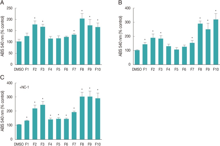
The toxicity of flavonoids F1 through F10 in glial cells was determined using the MTT assay. We observed that compared with control cultures (0.1% DMSO), flavonoids F2, F3, F7, F8, F9, and F10 (50 µM) induced an increase in cell viability in uninfected glial cell cultures (Fig. 1A, B) after 24 hr and 48 hr of treatment. Additionally, an increase in cell viability was observed in NC-1 infected cultures pretreated with all flavonoids (Fig. 1C). These effects were more pronounced in the cultures treated with flavonoids F2, F3, F8, F9, and F10.
Effect of flavonoids on N. caninum infection in glial cell cultures
To investigate whether a 24 hr pretreatment of glial cells with flavonoids interferes with NC-1 infection, we quantified the number of tachyzoites and the proportion of glial cells with parasitophorous vacuoles in infected cultures with or without flavonoid pretreatment. Fig. 2A shows the numbers of NC-1 tachyzoites present in cell lysates 24 hr after infection. The numbers of tachyzoites in cultures pretreated with flavonoids did not differ from those in cultures performed under control conditions (DMSO 0.1%). This may be because dead and alive tachyzoites did not differ morphologically, as observed under an optical phase microscope, and were counted together. However, analysis of NC-1 by immunocytochemistry and Rosenfeld staining showed that flavonoid pretreatment altered the proportion of glial cells presenting parasitophorous vacuoles. As shown in Fig. 2, approximately 64.4% (±9.2% SD) of the cells in cultures infected with NC-1 under control conditions (DMSO 0.1%) exhibited parasitophorous vacuoles. Additionally, F3, F6, and F9 significantly reduced the proportion of cells with vacuoles. However, flavonoids F1, F4, and F10 increased the proportion of cells infected with NC-1, and this difference was statistically significant.
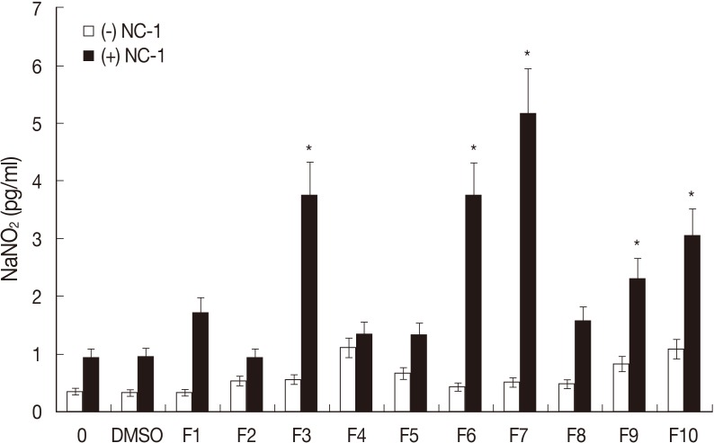
To investigate whether flavonoids and NC-1 infection interfere with glial cell functioning, the release of the trophic factor NO was evaluated. Nitrite levels in the culture media of control cultures (0.1% DMSO), from which NO levels were inferred, were very low (0.96 pg/ml) (Fig. 3), and no significant increase in nitrite levels was observed following the 50 µM flavonoid treatments. However, an increase in nitrite levels was observed in cultures exposed to flavonoids F3, F6, F7, F9, and F10 and infected with NC-1, with these levels reaching 3.75, 3.72, 5.17, 2.31, and 3.05 pg/ml, respectively.
DISCUSSION
Among the tested flavonoids, F1, F2, F3, F7, F8, F9, and F10 induced a significant increase in MTT metabolism in both glial cell cultures infected with NC-1 and uninfected cultures. MTT conversion primarily occurs via mitochondrial dehydrogenases and is dependent on respiratory chain activity [23]. Furthermore, increased activity of mitochondrial dehydrogenases in culture systems can be attributed either to an overall increase in metabolic activity or an increase in the number of MTT-metabolizing cells due to cell proliferation. Thus, it cannot be ruled out that flavonoids F1, F2, F3, F7, F8, F9, and F10 may have induced an increase in the metabolic activity of glial cells, particularly astrocytes, which are associated with the induction of microglial proliferation. Flavonoids F4, F5, and F6 all contain hydroxyl groups at positions 5 and 7 of the A ring, which may be linked to the biological response patterns observed.
Another pattern of glial cell responses resulting from flavonoid treatment and NC-1 infection was related to the proportion of cells with parasitophorous vacuoles. The biology of N. caninum is characterized by rapid infection and spreading in the cytoplasm of host cells. In glial cell cultures, it has been determined that these parasites replicate in approximately 6 hr [7,8]. Apparently, pretreatment of glial cells with flavonoids F2, F5, F7, F8, and F10 did not affect the proliferation of this parasite. The cultures pretreated with flavonoids showed a proportion of parasitophorous vacuoles similar to that observed under control conditions. However, pretreatment of glial cells with flavonoids F3, F6, and F9 clearly reduced the proportion of infected cells presenting parasitophorous vacuoles containing NC-1. These molecules all present hydroxyls groups at the 3' and 4' positions of the B ring of their structures, forming a catechol nucleus, which has been attributed to toxicity in different biological systems. This characteristic may confer a toxic effect of flavonoids on NC-1 if this structure remains intact and biologically active at the time of infection. Moreover, this result may be due to an indirect effect of flavonoids on glial cell responses that interfered with the infection and multiplication of parasites in these host cells. In the mammalian CNS, NO can be produced via a free radical generation system. This system is absent in glial cells, but NO production can be stimulated through the conversion of arginine to citrulline via inducible NO synthase. Expression of this enzyme has been detected in cultures of glial cells and astrocytes in neuropathological samples [27]. In the present study, a significant increase in the accumulation of nitrite, the stable form of NO, in the culture medium of cells treated with flavonoids was observed. However, this induction of NO production was observed in cultures infected with NC-1 when they were pretreated with flavonoids F3, F6, F7, F9, and F10. These findings suggest that there was a relationship between NO production and the prevention of NC-1 infection in the glial cell cultures.
Taken together, our findings clearly show that glial cells (astrocytes and microglia) respond differently to flavonoids exhibiting distinct degrees of hydroxylation and that these compounds induce glia activation, which interferes with N. caninum infection. Among the flavonoids tested and the evaluation parameters adopted in this study, F3 (3',4'-dihydroxyflavone), F6 (luteolin, 3',4',5',7-tetrahydroxyflavone), and F9 (quercetin, 3,3',4',5,6-pentahydroxyflavone) presented the most promising anti-parasitic potential. These drugs both induced glial cell activation and secretion of NO and inhibited N. caninum parasitophorous vacuole formation.