INTRODUCTION
Giardia is a unicellular parasite that infects a wide range of vertebrate hosts, including humans [1]. Six species have been identified for this intestinal parasite. To date, Giardia lamblia (syn. G. duodenalis or G. intestinalis) is the only species associated with human infections [2]. Infection is usually transmitted through ingestion of infective cysts, the environmental stage [1]. Infection occurs worldwide, but mostly affects populations in the developing countries [3]. Infection is commonly asymptomatic but mild to moderate self-limiting diarrhea occurs in some cases [3]. In other cases, diarrhea may be severe, prolonged, and even threatening to life [2,3]. According to a recent study, G. lamblia has 7 genotypes, frequently referred to as assemblages, named alphabetically from A to G [4]. Although assemblage E has been isolated recently from human feces [5], most of infections occurring in humans have been attributed to genotypes A and B [4,6]. Due to the great genetic diversity between the 2 genotypes, assemblages A and B have been considered by a number of investigators as 2 distinct species [7].
The diagnosis of Giardia infection relies entirely on laboratory diagnosis. Examination of feces with classical wet mount microscopy is frequently employed as a rapid, cheap, and simple method [8,9]. However, microscopy offers a low sensitivity and depends to a great extent on the skill of the person who is carrying out the test. Several antigen detec tion kits, frequently recognized as “coproantigen” kits are commercially available cost-effective diagnostic methods, alternative to microscopy [9,10]. However, these kits have reported lower specificity and sensitivity than that obtained with the PCR-based detection tools. PCR is a more important method not only for epidemiological studies, but also as a diagnostic tool for low-density Giardia infections [11].
Despite several PCR-based diagnostic assays which have been described [12-17], the implementation of the majority of these assays into microbiology laboratories, particularly in resource-poor countries, is still challenging. This may be related to a number of reasons. First, the majority of these assays rely on multi-step procedures for Giardia DNA extraction and subsequent PCR amplification. Second, most of the assays lack standardization and proper clinical evaluations. Finally, for poor countries where Giardia infection predominates, PCR is still considered an expensive technique in comparison to the conventional diagnostic methods.
In this study, we aimed to develop a simple, internally-controlled, and cost-effective PCR-RFLP assay for detection and characterization of G. lamblia clinical isolates in human feces. The assay performance was investigated in comparison to microscopy and a rapid immunoassay with fecal samples collected from asymptomatic Giardia human infections. The agreements between PCR assay results and that of the rapid immunoassay, microscopy, and the reference PCR were also investigated.
MATERIALS AND METHODS
Giardia strains
The WB-assemblage A (ATCC® 50803) and GS-assemblage B strains (ATCC® 50581) were purchased from the American Type Culture Collection (Rockville, Maryland, USA) and grown in the laboratory using a modified TYI-S-33 medium as described previously [18].
Fecal samples
A total of 563 fecal specimens were collected from those submitted to the Microbiology Department at 2 public hospitals at Taif region, Western Saudi Arabia, for routine parasitological examinations. The initial 91 samples were subjected to parasitological examinations with composite gold standard tests comprising of iodine-stained microscopy method [9], RIDA® Quick Giardia rapid kit (R-Biopharm, Darmstadt, Germany), and small subunit (SSU) rRNA PCR [19]. According to the test results, 4 groups of control samples were prepared and used while estimating the diagnostic performance of the assay. Group-1 included 15 samples diagnosed as Giardia-positive by the 3 methods. Group-2 involved 22 samples described as Giardia-negative with microscopy but were positive for Giardia coproantigen and DNA. Group-3 contained 9 samples that were Giardia-negative by microscopy and rapid immunoassay but were Giardia DNA-positive with the reference PCR. Lastly, group-4 comprised of 45 Giardia-negative samples with 3 individual assays. Microscopic examination of these Giardia-negative samples revealed Entamoeba histolytica/dispar, Dientamoeba fragilis, Blastocystis hominis, and Hymenolepis nana in 7, 3, 4, and 2 samples, respectively.
Subsequently, the remaining 472 samples were used to validate the diagnostic PCR assay. Fresh specimens were examined by the routine wet mount microscopy followed by the rapid immunoassay test within 2-3 days after collection. The remainder of each specimen was adequately labeled and kept without any preservative at -20˚C till PCR examination.
DNA extraction protocol
Nucleic acid was directly extracted from whole fecal samples, cultured Giardia trophozoites, Giardia cysts-spiked feces, or purified Giardia cysts suspension using the QIAamp® Stool Mini Kit (Qiagen, Valencia, California, USA) following an amended kit’s protocol [20]. Briefly, extraction was performed using 200 µl of feces and eluted into 50 µl of the elution buffer. The lysis temperature was raised to the boiling point for 10 min, and the incubation time of the InhibitEX tablet was extended to 5 min. Also, a pre-cooled ethanol was used for nucleic acid precipitation.
PCR amplification
Approximately 455 bp of Giardia glutamate dehydrogenase gene (gdh) sequence was amplified using 2 previously described specific primers; GDHeF and GDHiR [21]. Amlifications were carried out in 96-well TechneTM TC-4000 thermal cycler (Techne, Cambridge, UK). Following optimization experiments, reactions were set up with 10 pmol of each primer, 1 µl of DNA extract, 1X Go Taq® green buffer, 0.2 mM of each dNTP (Bioline, London, UK), 1.5 mM MgCl2, 1% DMSO, and 1 U of GoTaq® HotStart (Promega, Madison, Wisconsin, USA) as final concentrations.
Reaction of 20 µl volumes were prepared and run with a touchdown PCR protocol as follows: Initial denaturation was set up at 94˚C for 4 min, followed by 10 cycles of the touchdown stage with denaturation at 94˚C for 30 sec and annealing temperature at 67˚C for 30 sec. Then, the annealing temperature was decreased to 57˚C by 1˚C per cycle for the first 10 cycles. Extension temperature used was 72˚C for 30 sec and the subsequent 30 cycles were as follows; denaturation at 94˚C for 30 sec, annealing at 57˚C for 30 sec, and extension at 72˚C for 30 sec. A final extension step at 72˚C for 7-10 min was included.
PCR products were analyzed on ethidium bromide-stained 1-2% agarose gels (Sigma, Dorset, UK) in an in-house electrophoresis tank. Gels were viewed by UV illumination and photographed using a gel documentation system (Kodak, Rochester, New York, USA). The GeneRulerTM 100 bp DNA molecular weight marker (Fermentas, York, UK) was used as the size standard in gel electrophoresis.
Construction of PCR amplification controls
PCR reaction was set up with 1 µl of DNA extract of cultured WB strain. The PCR product (i.e., amplicon) was gel-purified with YORBIO Gel/PCR DNA Purification Kit (Yorkshire Bioscience, York, UK) and subsequently cloned into pCR®4-TOPO vector (Invitrogen, Paisley, UK) following the standard procedures [22]. The plasmid was then propagated in Escherichia coli (Top10) and subsequently purified with the Wizard Plus Miniprep isolation kit (Promega, Leiden, Netherlands) prior to sending for bidirectional sequencing (Eurofins MWG, Germany). Stocks of the plasmid construct were prepared for subsequent PCR reactions as an external control (EC).
Based on the retrieved EC sequence, an internal control (IC) of ≈ 300 bp was constructed through deleting ≈ 155 bp between the flanking primer sequences using the same approach that we have practiced in an earlier study [23]. Briefly, a forward primer (AGA AGC TTC TCA CAG GCA AGA ACG TCA A) and a reverse primer (ATA AGC TTA TGA CCT CGT TGT CGG ACT T) were manually-designed with HindIII restriction enzyme recognition sequence plus 2 additional nucleotides incorporated at the 5´ terminus of each. An inverse PCR was set up and as a result, an amplicon of ≈ 4.2 kb was produced. The amplicon was gel purified, subjected to restriction diggestion, and religated on itself forming plasmid with the new construct. Plasmid was propagated in E. coli (DH5α), and stocks of DNA was prepared as described above.
Optimum concentration of IC
Serial dilutions of EC plasmid DNA (1 ng-0.05 fg/µl) were prepared, subjected to PCR amplifications, and the lowest concentration with amplicon on gel was defined as the lower detection limit (LDL). Ten-fold serial dilutions of IC plasmid DNA (1 ng-0.1 fg/µl) were prepared and co-amplified with the EC plasmid DNA with concentration equal to the LDL, in 1 tube as a duplex PCR. The lowest dilution, consistently detectable with the primary PCR target on gel, was defined as the optimum concentration of IC.
Cyst purification, counting, and seeding experiment
Three G. lamblia cyst-positive fecal samples were pooled, concentrated, and purified initially with modified formol-ether concentration [9] and then by the sucrose density-gradient centrifugation techniques as previously described [24]. Cysts were counted under the microscope with a modified Fuchs-Rosenthal counting chamber (Hausser Scientific, Horsham, Pennsylvania, USA) following the manufacturer’s protocol. Suspensions of PBS, 1 ml each, with ≈ 106 of Giardia cysts were prepared, labelled, and stored at -20˚C till usage.
Estimation of PCR assay performance
Decimal serial dilutions of 106 cysts-spiked 200 μl-aliquot of Giardia-free fecal material were prepared and subjected to DNA extraction. The retrieve DNA extracts were amplified, and the lowest dilution that showed product on the gel was defined as the LDL.
Restriction digestion
Amplification products of 28 Giardia-positive samples, including samples with discordant results with microscopy and the immunoassay were gel-purified. In 20 µl volume reactions, ≈ 1 µg of the purified DNA was subjected to restriction digestion with 2U Nla IV (Roche, Burgess Hill, UK) in the presence of 1×reaction buffer. Digestion was carried out at 37˚C for 1-3 hr. Restriction digest profiles were analyzed on 2% high resolution grade agarose (Fisher Biotech, Loughborough, UK) stained with ethidium bromide and analyzed according to the restriction patterns described elswhere [21].
Ethical considerations
This research was carried out following an approval gained from the Local Directorate of Health and from the ethics committee of Taif University, Saudi Arabia. Permissions were also taken from patients attending hospitals to use their biological materials during the specified research period.
Statistical analysis
Data were collected and statistically analyzed with SPSS 17.0 software (SPSS Inc., Chicago, Illinois, USA). The diagnostic sensitivity (Se), specificity (Sp), positive predictive value (PPV), and negative predictive value (NPV) were assessed using chi-square tests against the combined gold standard test results. The agreement of the diagnostic assay results with the other tests was done with Kappa statistics (κ). Confidence intervals (CI) of proportions (95%) were calculated based on binomial distribution. The gdh copy number per 1 nanogram of plasmid DNA was calculated following a previously described equation [25].
RESULTS
Constructed plasmids
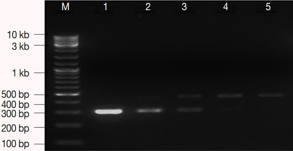
Two recombinant plasmids were constructed in the current study. Table 1 shows a number of important features for the 2 plasmids. The lowest concentration of the EC plasmid DNA that was successfully amplified and detected on gel, as a sole target of amplification, was 0.5 fg which corresponds to ≈ 100 copies of the gdh target gene locus. The estimated optimal concentration of IC DNA was 0.9-3.6 fg per reaction (Fig. 1). Stocks with the described concentrations were prepared, aliquoted, carefully labeled, and used as an EC and IC for the PCR assay in the subsequent amplification reactions.
Analytical performance of assay
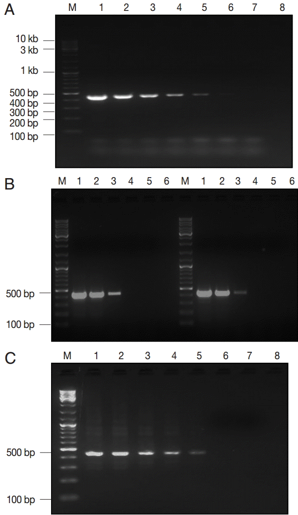
Three estimates of LDL of the assay were described according to the type and purification degree of the template DNA. The assay detected a minimum of 0.5 fg of EC plasmid DNA, corresponding to ≈ 100 copies of the gdh gene (Fig. 2A). Moreover, concentration of ≈ 2 pg of genomic DNA (WB and GS strains) was the LDL, which matched to ≈ 12 copies of the Giardia genome (Fig. 2B). Lastly, Giardia DNA was successfully extracted and subsequently amplified from all spiked samples down to ≈ 100 cysts per extract (200 μl), corresponding to ≈ 2 cysts per PCR reaction (Fig. 2C).
Diagnostic performance of assay
The assay successfully amplified DNA sequences from all Giardia-positive control stool samples except 3 samples which belonged to group-3. Running an additional PCR round with 1 µl of the PCR product as a template, the primary target DNA was identified in these 3 samples. Importantly, no amplification was detected for G. lamblia specific DNA in any of the Giardia-negative control samples (n=45). Equally important, the IC target DNA was successfully amplified in all samples (Fig. 3). Based on these results, the PCR assay was found to exhibit Se, Sp, NPV, and PPV of ≈ 93%, 100%, ≈ 94%, and 100%, respectively (Table 2).
Application to clinical samples
G. lamblia DNA was exhibited in 5.9% (28/472) of samples by the diagnostic PCR assay. Giardia coproantigen was identified in 5.2% (25/472) of samples by RIDA® Quick kit. The reference PCR assay demonstrated G. lamblia DNA in 6.1% (29/472) of samples. With iodine-stained microscopy ≈ 8% of samples were positives for Giardia and/or other intestinal parasites. G. lamblia cysts were demonstrated in 3.6% (17/472) of the examined feces. Out of these 17 Giardia-positive samples, 12 were sole Giardia infections while the remaining 5 samples were found mixed with other intestinal parasites. Giardia cysts were concomitantly found in 2 samples with E. histolytica/dispar cysts, in 2 samples with B. hominis cysts and in 1 sample with D. fragilis. Lastly, B. hominis, E. histolytica/E. dispar, H. nana, and D. fragilis were also demonstrated as sole infections in 8, 7, 2, and 1 Giardia-negative sample, respectively.
Aggreament with other test results
Table 3 shows the percent of agreement between PCR assay results and that of the other tests. All Giardia-positive samples by microscopy were also positives by the immunoassay and 2 PCR assays. All Giardia-positive samples by the immunoassay were also positive by 2 PCR assays, apart from 1 sample. No significant PCR inhibition was noticed in all Giardia-negative samples.
G. lamblia genotypes
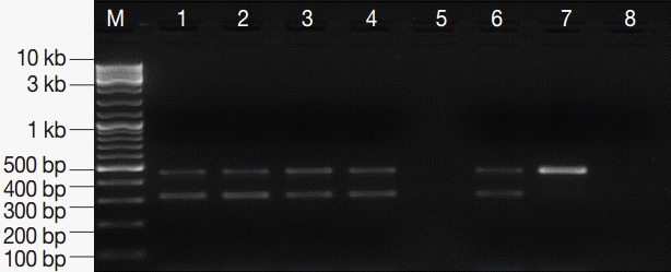
DNA products of all 28 Giardia-positive samples by the developed assay were subjected to restriction digestion by Nla IV, and 12 restriction profiles were G. lamblia genotype A while the remaining 16 profiles were G. lamblia genotype B. None of the examined samples showed mixed genotypes.
DISCUSSION
In earlier studies, a variety of genetic loci including (SSU) rRNA, gdh, triose phosphate isomerase, β-giardin, and elongation factor 1-α have been used as the target gene loci for Giardia PCR assays [7]. The (SSU) rRNA gene sequence is short and highly conserved among the major G. lamblia genotypes, thus it has been mainly used in PCR detection assays [19]. Conversely, the other genes have frequently been used in detection and characterization studies [12-17].
In the current study, we developed a simple PCR assay for identification and subsequent discrimination of Giardia clinical isolates in human feces based on specified gdh gene sequence. A simple method was selected to extract and purify Giardia DNA directly from feces without any sample preparatory steps. The whole method was carried out in 1-2 ml scale, hence permitting the use of inexpensive table-top microfuges and heating blocks that are available at most of the basic laboratories. Extraction of 24-sample batch was carried out in ≈ 120 min. The commercial kit-based extraction method has been proved useful for extraction of more than 1 protozoan DNA directly from feces [20].
Being an enzymatic reaction, PCR is amenable to inhibition by substances in feces or accidentally brought into reactions [26]. PCR inhibition gives rise to false negative results that are not acceptable in clinical laboratory settings. Thus, we validated our PCR assay with an IC with a minimal additional cost. The concomitant use of IC with the primary amplification target in a duplex PCR worked properly without causing alteration to the diagnostic efficiency of the assay. No significant inhibition was noticed for all DNA retrieved from feces which reflects the high efficiency of the extraction protocol adopted in the study, consistent with our previous findings [20].
The analytical sensitivity of the assay was demonstrated, and ≈ 2 pg of genomic DNA was sufficient for detection, in agreement with the original study [21]. Read et al. [21] reported identical sensitivity but for a semi-nested assay with DNA purified from a cultured strain p1c10, representative of G. lamblia, genotype A. Although nested or semi-nested PCR assays help in increasing the sensitivity and specificity of the target sequence detection, assays with that formats also cause more elevation of assay cost, duration, and increase the risk of PCR carry-over contamination [27]. Also in this study, another estimate for the analytical sensitivity which reflected to some extent the efficiency of both the extraction and amplification steps, was undertaken in this study. Assuming that the cyst count seeded in stool aliquots was precise and the DNA extraction was carried out from all cysts with equal efficiency, ≈ 2 cysts were sufficient for identification. It is important to announce that, in view of the estimated assay sensitivity and Giardia cysts excretion rates that have been reported in human infections [28], this sensitivity is considered acceptable.
The diagnostic performance of the assay was addressed using high quality control samples that were prepared in the study. A sensitivity of 93% and a specificity of 100% were demonstrated by the assay. The 3 Giardia-positive control samples that could not be picked up by the assay were assumed to be of low parasite loads. The exhibited outstanding assay specificity may be related to the high specificity of primers, the usage of the touchdown protocol, and inclusion of 1% DMSO in the reaction, as previously stated [29,30]. Subsequently, the assay was validated on a panel of stool samples with blinded results. Giardia infection was detected in the study population with a prevalence rate of 5.9%, slightly lower than that described previously. According to earlier reports, Giardia has been found in 8.5-17.0% of population from Taif [31,32]. Moreover, prevalence rates of 3-9% have been described in other Saudi populations [33,34]. Much higher rates have been recorded for Giardia infections in developing countries outside Saudi Arabia [35]. Certainly, these prevalence variations among studies may be explained by the differences in the study design, target population, sampling size, and the adopted parasitological examination [36].
The restriction digestion profiles of 28 Giardia DNA-positive samples were successfully analyzed. Both genotypes A and B were described in the study population. The identification of Giardia clinical isolate is far important not only for epidemiological studies but for infection control programs as well. The correlation between each genotype and clinical picture of Giardia infection has been investigated but still uncertain. While a number of investigators have linked genotype A with mild infection and genotype B with severe infection [37], others have found the above correlations insignificant [28]. These associations could not be investigated in this study because the clinical data extracted from hospital records were not sufficient.
Like any new diagnostic tool, evaluation of our PCR assay has to be carried out continuously. A larger prospective clinical study including samples from various geographic locations covering both endemic and non-endemic regions has to be done in the future. Moreover, further measures for improving the assay have to be searched for in view of the rapidly evolving PCR technology. The assay could be improved more in the future by inclusion of more detection targets of clinically-related and epidemiologically relevant enteric pathogens (viruses, bacteria, and/or parasites)-attributed diarrhea. Moreover, the simple kit-based extraction protocol and the single-round PCR amplification adopted in the PCR assay make the assay highly amenable for partial or complete automation with subsequent reduction in cost, time, and human error.
In conclusion, we developed a diagnostic PCR-based assay coupled with restriction digestion polymorphism for identification of Giardia clinical isolates in human feces. The assay was proved simple, relatively cost-effective, highly specific, adequately sensitive, and reliable. The assay offers a great potential to be used as a diagnostic test in clinical laboratories, particularly in developing countries. Future studies are required for further evaluation, and future measures have to be sought for more improvement of the assay in view of the rapidly progressing PCR technology with more reduction of PCR reagents and machines.