INTRODUCTION
Zoonotic diseases in small mammals are a risk to humans who work or conduct recreational activities in field environments. During the fall period, typical zoonotic diseases in the Republic of Korea (ROK) are scrub typhus, hantaviruses, and leptospirosis [1]. They annually occur and cause acute, febrile illness to humans. Scrub typhus especially became increasingly important. According to the Korea Centers for Disease Control and Prevention (KCDC), chigger mite distribution has expanded from the south to central Korea. The number of scrub typhus patients increased from approximately 200 cases in the early 1990s, to >10,000 cases in 2013 [1]. Known species of chigger mites include Leptotrombidium pallidum, L. scutellare, L. palpale, L, orientale, L. zetum, Neotrombicula gardellai, Euchoengastia koreaensis, Neotrombicula japonica in the ROK. L. pallidum is the predominant species in the central region, and L. scutellare in the southern region [2]. In the world, there have been more than 20 antigenically distinct strains of scrub typhus reported [3]. The prototype Kato, Karp, and Gilliam strains were classified into a higher virulence group for humans. The Boryong type, which is a major strain in ROK, was classified into a lower one [4].
Recent studies emphasized the relationships between a warming climate and distribution of primary scrub typhus vectors [5,6], as well as detection of O. tsutsugamushi positive chigger mites, including new species as potential vectors of O. tsutsugamushi [7]. Therefore, this investigation focused on scrub typhus and other zoonotic diseases among small mammals and associated O. tsutsugamushi positive chigger mites. This information will provide baseline data for future investigation and information for development of zoonotic diseases mitigation strategies.
MATERIALS AND METHODS
Collection site
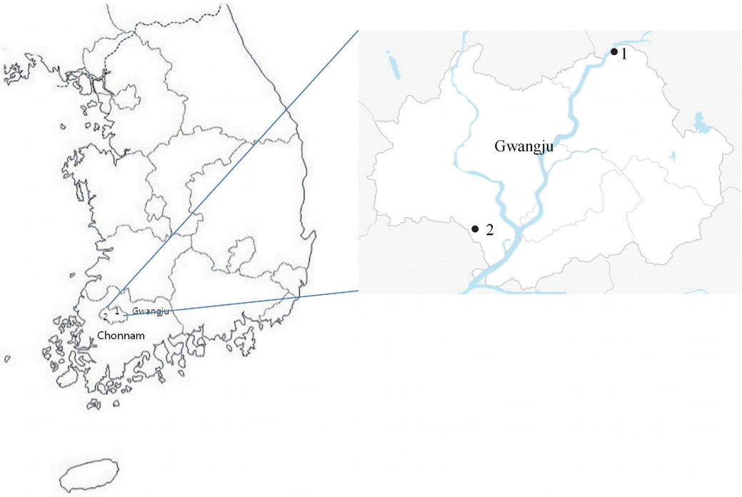
From September 2014 to August 2015, small mammals were collected using Sherman live traps (3"×3.5"×9", USA) in 2 regions (Gwangsangu and Bukgu) of Gwangju Metropolitan Area (MA), ROK. Collection sites were just within the city perimeter. The 2 regions where traps were installed were in a similar environment. Both included 5 types of locations. The locations were fallow ground, a ridge between rice fields, a boundary between forest and field, around tombs, and around water. Traps were baited with biscuits covered with peanut butter and set up in the late afternoon, and collected from 8-10 a.m, the following morning. Gwangju MA surrounded by Chonnam Province is a densely populated area (Fig. 1).
Collection of rodents and chiggers
Traps positive for small mammals were numbered, placed into secure shipping containers, and then transported to the Health and Environment Research Institute of Gwangju MA. Small mammals were euthanized using chloroform (Merck, West Point, Pennsylvania, USA) soaked cotton (1×1 cm). Then, blood was centrifuged at 3,000 rpm for 20 min, and serum was separated and maintained at 4˚C. Indirect immunofluorescence assay test (IFAT) and passive hemagglutination assay (PHA) were performed 24-36 hr after the small mammals were euthanized for antibody detection. The carcasses of the small mammals were hung over a glass bowl containing water to harvest chiggers, and chiggers were collected the following day.
Detection of O. tsutsugamushi, hantantaviruses, and Rickettia spp. antibodies in small mammals
A total of 10 μl of sera from each small mammal was used for serial dilutions of 1:16, 1:32, 1:64, 1:128, 1:256, 1:512, 1:1,024, and 1:2,048 in PBS (pH 7.2). Diluted sera were deposited on an antigen spot slide, incubated at 37˚C for 30 min in a humidified chamber, and then washed as in step 2. First, the slide was washed for 3 min with PBS, and second, washed for 3 min with distilled water to remove PBS salt. A total of 25 μl fluorescein isothiocyanate (FITC)-conjugated goat anti-mouse IgG (Sigma, St. Louis, Missouri, USA) was added to a spot slide, and the slides were incubated at 37˚C for 30 min in a humidified chamber. The slides were washed for 3 min with PBS, distilled water, and then air-dried. After mounting medium (Sigma) was added to a spot slide, and covered with coverslip, the slides were examined for specific spots using a fluorescence microscope (Carl Zeiss, Oberkochen, Germany). A cutoff titer of 1:16 was used to identify seropositivity. Antigen spot slides for O. tsutsugamushi, hantaviruses, and Rickettsia spp. were provided by KCDC.
Detection of Leptospira spp. antibodies in small mammals
Genedia Lepto PHA (Green Cross, Seoul, Korea) kit reagents were used for detection of leptospirosis by PHA. A total of 25 μl serum from each small mammal was added to a 96-well plate, and diluted 1:80 in a dissolved solution of Genedia Lepto kit. A total of 75 μl of sheep blood containing red blood cells sensitizied by Leptospira spp. was placed on a diluted serum for agglutination assay. Ag-Ab agglutination reaction in 1: 80 tested positive for leptospirosis.
Detection of O. tsutsugamushi in chigger mites by PCR
The method used by Ree et al. [8] was applied for the detection of O. tsutsugamushi from individual chiggers. Individual chiggers were placed on a glass slide with PBS 10 μl. The chigger’s internal contents were squeezed out with 2 fine pins and observed after suspension with 50 μl PBS under stereomicroscope (Carl Zeiss). The chigger exoskeleton was mounted with polyvinylalcohol medium (Bioquip, Gardena, California, USA) and identified by Ree’s fauna key [9]. Because it was difficult to perform with all chiggers, 30 chiggers per small mammals were used.
DNA was extracted from 20 μl of chigger suspension using G-spin total DNA extraction kit (cat. no. 17046; Intron Biotechnology, Seoul, Korea). 200 μl of lysis buffer and 10 μl of proteinase K solution (20 mg/ml) was added to 20 μl of chigger suspension. The lysate was incubated at 56˚C on a heating block for 30 min. After lysis, 200 μl of binding buffer was added. Then, the mixture was incubated at 70˚C for 5 min. The mixture was applied to the spin column, and centrifuged at 13,000 rpm for 1 min. After discarding the filtrate, washing buffer A was added to the spin column and centrifuged at 13,000 rpm for 1 min. After discarding the flow-through, the column was placed into a 2.0 ml collection tube. Washing buffer B was added to the spin column and centrifuged at 13,000 rpm for 1 min. After discarding the flow-through, placed column into a new 1.5 ml collection tube, and a total of 50 μl of elution buffer was added directly onto the membrane. After incubating for 1 min at room temperature, it was centrifuged for 1 min at 13,000 rpm to elute. DNA extract was stored at -20˚C until amplification.
PCR was performed as INNOPLEX TSUTSU detection kit (cat. No. IPC10040; Intron Biotechnology). The kit was designed using primer sets to detect the 475 bp fragment gene encoding the 56 kDa antigen of O. tsutsugamushi. The first PCR was performed with 2 μl of DNA extract and 18 μl of distilled water treated with diehyl pyrocarbonate (DEPC; Gendepot, Barker, Texas, USA) in the first PCR premix tube. PCR conditions were as follows: initial denaturation at 94˚C for 5 min; 40 cycle at 94˚C for 30 sec, 58˚C for 30 sec, 72˚C for 40 sec; and final elongation at 72˚C for 5 min using a Geneamp 9700 Biosystem (ABI, Foster City, California, USA). The second PCR was performed with 2 μl of the first PCR product and 18 μl of distilled water with DEPC in the second PCR premix tube. PCR conditions were as follows: initial denaturation at 94˚C for 5 min; 30 cycle at 94˚C for 30 sec, 58˚C for 30 sec, 72˚C for 40 sec, and final elongation at 72˚C for 5 min using a Geneamp 9700 Biosystem (ABI). The final PCR products were evaluated by 1.5% agarose gel electrophoresis containing ethidium bromide.
The nucleotide sequence homology was aligned with sequence of previously published O. tsutsugamushi in Genbank by the BIOEDIT software program. The phylogenetic tree was generated by the neighbor-joining method (MEGA 6.0).
RESULTS
Small mammal collection
A total of 172 small mammals were collected. Apodemus agrarius was the most frequently collected 158 (91.8%), followed by Myodes regulus 8 (4.6%) and Crocidura lasiura 6 (3.5%). Overall, monthly mean trap rate was 14.3% (range 7-26%) (Table 1). For all periods, the trap rate for A. agrarius was higher for fallow ground (34.2%), followed by around water (29.1%), a boundary between forest and field (17.7%), around a tomb (13.9%), and a ridge between grain fields (5.1%) (Table 2).
Seropositive rates for O. tsutsugamushi, hantaviruses, Rickettisia spp., and Leptospira spp. in small mammals
Blood samples were obtained from 156/172 (90.7%). A total of 41/156 (26.3%) were seropositive for O. tsutsugamushi followed by small mammals positive for hantaviruses (24/156, 15.4%), Rickettsia spp. (22/156, 16.7%), and Leptospira spp. (2/172, 1.3%). Especially, tested 12 rodents were all positive for O. tsutsugamushi in October 2014. The prevalence for 4 vector-borne (deletion) infectious diseases was confirmed all the year round in Gwangju MA. Total 21 cases of mixed infections were confirmed in A. agrarius (Table 3).
Seasonal distribution of chiggers and detection of O. tsutsugamushi by PCR
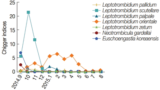
A total of 2,772 chiggers were collected from 158 A. agrarius (Table 4). L. scutellare 579 (54.1%) was the most commonly collected chiggers, followed by L. orientale 281 (1.8%), E. koreaensis 94 (8.8%), L. palpale 46 (4.3%), L. pallidum 36 (3.3%), and L. zetum 7 (0.7%). The majority of L. scutellare were collected during the autumn (September-November, mean 5.6-21.4), while L. orientale was collected in late autumn (October, mean 1.7) through spring (May, mean 3.0). A high number of E. koreaensis was collected in fall (September, 6.8), but infrequently during other months (Fig. 2).
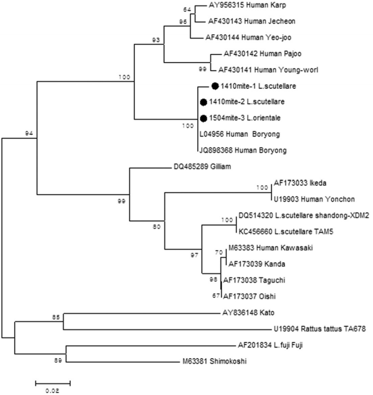
The distribution of chiggers by species of small mammals was 7 chigger species on A. agrarius, 4 species on M. regulus, and no chiggers on C. lasiura (Table 5). A total of 3 chiggers (0.24%) were positive for O. tsutsugamushi (Table 5). Each sequence (1,410 mite-1 L. scutellare, 1,410 mite-2 L. scutellare, 1,504 mite-3 L. orientale) showed homology of 90.1%, 93.2%, and 98.2% with the Boryong strain (accession no. L04956) (Fig. 3). Two L. scutellare collected from A. agrarius (September 2014), and 1 L. orientale from M. regulus (April 2015) were confirmed as positive for O. tsutsugamushi.
DISCUSSION
Seasonal serosurveillance of O. tsutsugamushi, hantavirues, Rickettsia spp., and Leptospira spp. in small mammals and a study on chiggers collected from small mammals were conducted. For all periods, A. agrarius was the most frequently collected species (91.8%). Capture rates of A. agrarius were high for fallow ground area (34.2%) and around water (29.1%), places which were unmanaged and covered with 1 m cyperaceae. There were always several puddles in the fallow ground area, and this area was more likely to be damp than other areas. C. lasiura and M. regulus capture rates were high for the boundary between forest and field areas and the area around tombs, which were located in a hillier section than the fallow ground area. There was few grass bush in these areas. There were differences in species distribution by areas.
As shown in Table 3, the prevalence of scrub typhus was 25.5% in A. agrarius, lower than in surveys performed in Gyeonggi province, central part of ROK [10-12]. We confirmed evidence of infection of various zoonotic diseases in A. agrarius and observed evidence of scrub typhus and hantavirues in M. regulus. Evidence of antibodies to hantaviruses and Rickettsia spp. implies that Gwangju MA area is not free of other zoonotic diseases.
In chigger mite’s distribution, L. scutellare was the predominant chigger from A. agrarius in Gwangju MA and was similar to the results at Chonnam province [13-15]. Meanwhile, L. pallidum was the predominant species in central parts of ROK [16-18]. As shown in Fig. 2, there was seasonal difference of chigger mite species collected from A. agrarius. L. scutellare was highest from October through November 2014, and high populations of L. scutellare may contribute to human transmission of scrub typhus in Gwangju MA area. L. orientale was highest in winter-spring seasons. This result indicates that the species of chigger mites could be affected by temperature and humidity.
Strains of scrub typhus show geographical variation. In ROK, the Boryong strain is known to be the predominant type in vectors and humans [3]. Our results showed that the 3 chiggers positive for scrub typhus were all Boryong strain. In our study, it was found that small mammals were the source of various zoonotic diseases, and L. scutellare contributed to high chigger indices during the fall period. To reduce risk of zoonotic disease in Gwangju MA, it is necessary to clear fallow ground near humans’ living space (cutting grass, filling puddles) for rodent control, and to provide adequate protection to farm workers (long sleeve clothes, boots). More information must be relayed about the risk of vector borne zoonotic diseases. Because information from 2 sites was limited, more data are needed over years regarding the relationship of disease transmission on humans, additional site selection, and distribution of gravid small mammals. In the future, our data will be helpful to provde prevention strategies against the resurgence of vector borne zoonotic diseases.