INTRODUCTION
A small-scale fecal examination subjected residents at a borderline village in North Korea (NK) and found high egg positive rates of Ascaris and Trichuris (Li et al., 2006). This suggests that NK may be an island of soil-transmitted helminthiases surrounded by the helminthiasis-free zones, South Korea (SK) and Northeast China. Those helminthiases are directly related with eating habits and food supply. During the last 10 years, the economic situation of NK has become worse, and most of the residents are facing difficulties of food supply. The difficulties of food supply mean massive starvations and eventually induce many diseases, which conform a vicious cycle of poverty and disease.
In the past, there was no difference in the infection status of helminthiases between South and North Korea (Kobayashi, 1928; Brooke et al., 1956). In recent 30 years, however, the situation of parasitic helminthiases has been changed greatly between South and North. Successful developments of economy and intensive control activities have successfully eliminated intestinal helminthiases in SK (Lee, 2005). Especially soil-transmitted helminths disappeared, but some foodborne parasites, zoonotic parasites, and malaria are still prevalent in SK (Korea Association of Health Promotion, 2004; Hong et al., 2006).
At present, ELISA is a standard diagnostic method for tissue-parasitic helminthiases in Korea, most of which are not detected by fecal examination (Cho et al., 1981; Yang et al., 1983; Kim et al., 1984; Cho et al., 1986; Hong, 1988). The ELISA detects specific serum IgG antibodies to 4 tissue-invading helminths; Clonorchis sinensis, Paragonimus westermani, Taenia solium metacestode, and sparganum (plerocercoid of Spirometra erinacei). These 4 helminthiases are slowly decreasing in SK now, but clonorchiasis is still prevalent forming wide endemic zones along big rivers. However, no information is available for them in NK at present.
The present study serologically examined sera of some residents and refugees from NK by ELISA to the 4 tissue-parasitic helminths. The present serology finding may provide information of helminthiases in NK, though the number of samples is limited.
MATERIALS AND METHODS
Subjected materials
We collected 137 sera (1 ml/person) of patients from a hospital in Cheongjin-shi, Hamgyeongbuk-do NK, under cooperation with staff of the hospital and also of Yanbian University, China in 2004. Most of the patients complained of common non-specific symptoms, such as fever, headache, nausea, abdominal pain, etc. The collected sera were delivered to Yanbian University and stored at -20℃, then transported to our laboratory in Seoul. In addition, we also collected 133 sera of female refugees who were living at a NK immigrant house in suburbs Seoul, SK in 2004. The NK subjects aged from 5 to 79 (median 39), and composed of 68 males and 202 females.
Serology by ELISA
Antigens of the tissue-parasitic helminths were crude extracts of C. sinensis and P. westermani, cystic fluid of T. solium metacestode and crude saline extract of spargana. The antigens were prepared in the ELISA laboratory of the Institute of Endemic Diseases, Seoul National University College of Medicine. Negative control sera, which showed no reaction to any of the 4 antigens, were collected from healthy students. Positive control sera were selected from single antibody positive patients who were diagnosed by routine ELISA.
To determine IgG antibodies to antigens of C. sinensis, P. westermani, T solium metacestode and sparganum in serum, ELISA was performed according to the standard method in our laboratory in 2005 (Lee et al., 2003). Each serum was tested twice and mean of the 2 absorbances were used. The antigens were diluted in carbonate buffer (pH 9.6) at 0.5, 0.5, 1.1, and 0.7 µg/100 µl protein concentration, respectively, and coated in wells of polystyrene microtitration plates (Costar, Cambridge, Massachusetts, USA) at 4℃ overnight. After washing, 1: 25 and 1: 100 diluted sera in phosphate buffered saline/0.05% Tween 20 (PBS/T, pH 7.4) were incubated for 2 hr at 37℃, respectively. After washing, 1: 24,000 diluted peroxidase-conjugated anti-human IgG (light and heavy-chain specific, Cappel, Aurora, Ohio, USA) in PBS/T was incubated for 2 hr at 37℃. After washing and taking advantage of tetramethylbenzidine (TMB, Ken-En-Tec Diagnostics, Taastrup, Denmark) reaction, absorbance was read at 450 nm using an ELISA reader (Molecular Devices Co, Sunnyvale, California, USA). Absorbance values of positive specific antibodies to C. sinensis, P. westermani, T. solium metacestode, and sparganum were over 0.250, 0.280, 0.290, and 0.240, respectively.
RESULTS
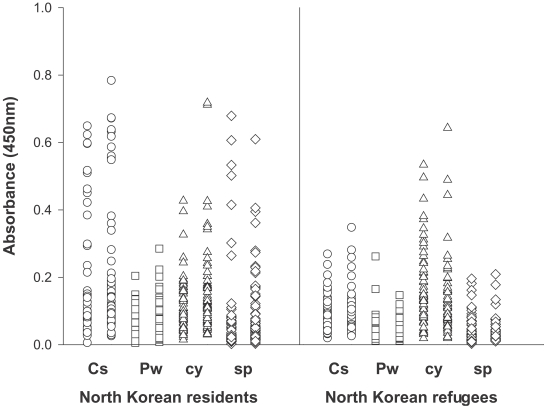
The ELISA of the 137 NK residents revealed positive rates of 19.7%, 8%, and 8% for antigens of C. sinensis, T. solium metacestode, and sparganum, respectively, but none were positive for the antigen of P. westermani. The total positive rate was 29.2%. By age, residents in their 50s showed the highest positive rate (Table 1 & Fig. 1). Of the 27 positive residents of clonorchiasis, 4 were duplicate positive to T. solium metacestode and 5 to sparganum. Most of the duplicate positive residents were in their age of 60s. The serology positive rates increased by age up to their 50s, but decreased in the 60s and 70s. Males were more positive than females, total 38.2% vs. 20.3%, and also in individual helminthiases. The NK refugees subjected in the present study were from 20s to 40s. They were positive to C. sinensis and T. solium metacestode by 3% and 10.5%, respectively (Table 1). There were no positive refugee subjects to P. westermani and sparganum.
Among a total of 270 samples, the antibody positive rates to the antigens of C. sinensis, T. solium metacestode, and sparganum were 11.5%, 9.3% and 4.1%, respectively. Some of them showed high absorbances (Fig. 1).
DISCUSSION
The present serological screening showed many serology positive individuals to the tissue-parasitic helminthiases, such as clonorchiasis, cysticercosis, and sparganosis in NK. The present data revealed 11.5% positive rate of clonorchiasis among all subjects and 19.7% among Cheongjin-shi residents. Therefore, we could confirm that clonorchiasis is prevalent in NK, considering sensitivity as 88.2% and specificity as 87.8% of the present ELISA system for diagnosis of clonorchiasis (Choi et al., 2003). More strictly, it is evident that clonorchiasis is highly prevalent among residents at Cheongjin-shi, Hamgyeongbuk-do, NK. Since clonorchiasis is a foodborne helminthiasis, which forms endemic areas along rivers and reservoirs, Cheongjin may be at the seaside margin of endemic areas of clonorchiasis in NK.
At present, clonorchiasis is endemic in SK and also in Northeast China (Korea Association of Health Promotion, 2004; Coordinating Office of the National Survey on the Important Human Parasitic Diseases, 2005). Since NK is located between the 2 endemic zones, we can simply presume that NK is endemic of clonorchiasis. Although we have no concrete data for distribution of intermediate hosts of C. sinensis in NK, ecological environment in NK must be suitable for transmission of C. sinensis.
None of the 270 sera was positive for P. westermani. Paragonimiasis is characteristically forming narrower endemic foci than clonorchiasis. Therefore, it is more difficult to find out an endemic area of paragonimiasis than that of clonorchiasis by this kind of small scale screening. Of course the present negative finding does not exclude the possibility of prevalence of paragonimiasis in Cheongjin-shi, NK, because of limited numbers and localities of the present study. The bioecology formed by mountainous natural environment in NK can support the possibility of enzootic transmission of Paragonimus. It must be a topic of further studies.
In the past, there were thousands of patients with cysticercosis, but new carriers are rarely found recently in SK. One previous serologic examination showed the positive rates of 2.1% among normal people and 4% among chronic epilepsy patients in SK (Kong et al., 1993). A serology paper of one hospital patients at Seoul observed 3.0% positive rate of cysticercosis (Lee et al., 2003). Compared to this, there are still newly recovered patients of cysticercosis and also infected pigs in northeast China (Hong ST, personal experience). The present finding of 8.0% serology positive rate of cysticercosis among residents in NK must be very high. This fact indicates that cysticercosis is also heavily prevalent at Cheongjin-shi, NK. This finding also means that T. solium is actively transmitted there. Poor general hygiene and insufficient medical care in NK are considered, and then cysticercosis must be the most serious helminthiasis of medical importance in NK. Since we have no information of the present subjected population, it is difficult to analyze further, but there must have been some clinical cases. We should concern cysticercosis when any control measure of parasitic diseases is planned in NK.
Sparganosis is distributed all over the world, and serologic examinations showed positive rates 1.6% to 3% in SK (Kong et al., 1994; Park et al., 2001; Lee et al., 2002; Lee et al., 2003). Sparganum is now enzootically transmitted among intermediate hosts and definitive hosts in SK (Sohn et al., 2005). The present serology positive rate of sparganosis is 8% among NK residents, which should be regarded as seriously high. Its positive rate was found higher among populations in a mountainous area than those in general population in SK (Park et al., 2001). Since most of the NK area is mountainous, the life cycle of sparganum is presumably well-maintained in NK. Some of the serology positive population in NK must be clinical patients of sparganosis.
Refugees from NK found serology positive rates of 3.0% to C. sinensis and 10.5% to T. solium metacestode. Their positive rate to T. solium metacestode was extremely high when it was compared with 2.1% of normal population and 4.0% of epileptic patients in SK (Kong et al., 1993). The positive rate of cysticercosis of the refugees was also a little higher than that of NK residents. Most of the refugees had stayed at neighboring countries for a certain period, such as China, Mongolia, Vietnam, Laos, and Thailand before they arrived at SK. During the stay at those countries, they might have had chances to contract infections not only of these food-borne helminthiases but of other diseases. Most of the regions on their migration route except for Mongolia are endemic of cysticercosis (Chung et al., 2005; Ikejima et al., 2005; Somers et al., 2006). The refugees stayed at the neighboring countries maximum up to 3 years, and their lives during the migration must have been very difficult for survival. We should consider cysticercosis or other infectious diseases for medical care of the NK refugees.
It is hard to generalize the present data through whole NK because the number of examined sera was too small and the locality was limited. Nonetheless, the positive finding of the tissue-parasitic helminthiases confirms their prevalence among North Koreans at present.
In conclusion, the present results confirm again that parasitic fauna of NK is same as that of SK, and that clonorchiasis, cysticercosis and sparganosis are prevalent in Hamgyeongbuk-do. We should concern tissue-parasitic trematodes and cestodes when any parasite control is planned for residents or refugees of NK. Since most of the helminths are transmitted by food, any food materials imported from NK must be inspected strictly. Also a warning is necessary for visitors to NK keeping from eating raw food there.