INTRODUCTION
Cystic echinococcosis (CE) also known as hydatidosis, caused by the metacestode of Echinococcus granulosus sensu lato, is a parasitic infectious disease of global health concern. Of 18 neglected tropical diseases (NTDs) listed by WHO [1], hydatidosis has endemicity to regions with prominent pastoral activities [2] impacting the poorest global societies thereby interrupting the efforts to achieve poverty alleviation and improvement in public health. Infectious diseases are posing serious challenges amid rapid human development resulting in emergence and reemergence of both old and new types. Main influencing factors are environmental and demographic changes which modify human-animal ecosystems. Such modifications lead to zoonoses of NTDs in particular which have lacked sufficient attention historically in context of international public health efforts leading to inadequate remedial measures [3].
Hydatidosis causes substantial economic losses in livestock and humans. Apart from being direct source of employment and income, livestock contributes critically in human nutrition. With rising global population and nutritional demand, reliance on livestock has increased manifold specially in developing countries. Complexities arise with heavy toll on livestock for the provision of products while compromising on animal health due to inefficient pasture management practices and production systems in resource poor communities [4].
Pakistan is considered as one of the endemic areas for CE [5] and other soil-transmitted helminths (STHs) with highest incidence rates for 8 NTDs [6]. Northern Punjab, Khyber Pakhtunkhaw (KP), Karachi and areas near Afghan border are at the highest risk of contracting this disease. CE is ranked as 4th most widespread helminth disease in Pakistan and there are possible chances of emergence of STHs (echinococcosis) in cities and their peripheries besides rural settings in coming decades [7].
A number of studies have reported mild to high prevalence rate of hydatidosis in Pakistan. Prevalence as high as 60.46% has been reported in buffaloes slaughtered at urban abattoirs of Punjab [8]. Pakistan is an agricultural economy; animal husbandry is widely practiced here in extensive conditions which is an established risk factor for hydatidosis [9]. Lack of proper sanitation, health and education facilities also correspond to making livestock a suitable reservoir for E. granulosus. Amidst such wide geographical presence and prevalence there is a dire need to have updated knowledge of current situation of hydatidosis.
This study would help in establishing the role of different domestic ungulates and assessing the transmission modalities of disease in Pakistan. Lack of such information may lead to inefficient control strategies and failure to eradicate hydatidosis.
MATERIALS AND METHODS
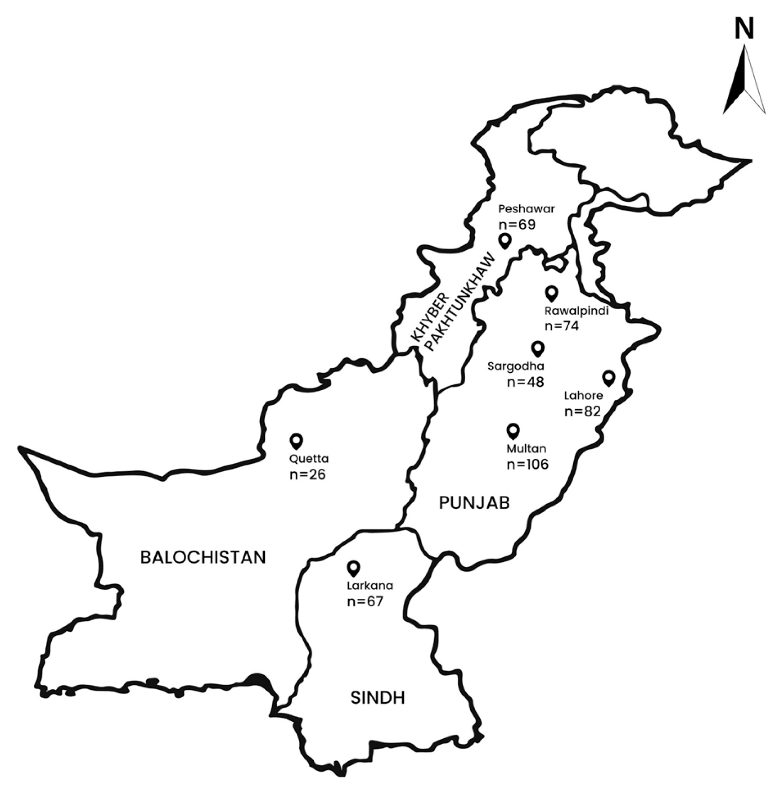
The study was carried out in 7 major cities of Pakistan: Quetta (Baluchistan province), Larkana (Sindh province), Peshawar (Khyber Pakhtunkhaw province), Multan, Lahore, Rawalpindi and Sargodha (Punjab province) (Fig. 1). The climate of these areas is predominantly hot with extended summers. These pastoral and agro-pastoral regions have large populations of domestic livestock. Livestock is one of the major sub-sectors of agriculture having a major role in economic sustainability and GDP of the country. Around 8 million households derive 35% of their income from livestock associated activities, forming backbone of rural economy [10].
Livestock slaughter is usually done at official abattoirs and local butcher shops in Pakistan. Moreover, animals from far flung areas are brought to cities for slaughtering. A total of 991 sheep, 1,478 goats, 1,602 cattle and 1,343 buffaloes were examined for presence of hydatid cysts during 2 years (January 2016–December 2018). Information like age (<1-≤2,>2-≤3,>3-≤5,>5 years) and gender (male, female) of the animal was taken at the spot through dentition pattern or asking the owner. Other data like condition of animal, previous disease history were also recorded.
Examination of slaughtered animals
Visceral organs of all slaughtered animals were examined for presence of hydatid cysts through palpation and visual inspection. All infected organs were brought to laboratory for further examination and cyst characterization. The infected organs were thoroughly examined to collect data for cyst number (<10 cysts and >10 cysts), form of cyst (single or multiple) and type of cyst (fertile, sterile, calcified/suppurative). Cysts were considered fertile if they contained protoscolices, sterile if they had no protoscolices but appeared viable and calcified if they were non-viable with degenerated lesions.
Statistical analysis
All data were subjected to descriptive statistics for estimating frequency and prevalence rates. Prevalence was taken as proportion of infected animals whereas intensity of infection was calculated as mean number of cysts in specified age groups, gender, season and infected organ [11]. Chi square statistic was employed, for delineating the role of different risk factors, through SPSS (version 20) at significance level of 0.05.
RESULTS
A total of 5414 animals (sheep, goats, cattle and buffaloes) were examined during the study period for determining the prevalence of hydatid cysts. Highest prevalence was found in buffaloes (11.9%) followed by sheep (11.5%), cattle (8.9%) and goats (3.6%). Overall prevalence of hydatidosis for all animal species was 8.71%. Chi square statistic was significant (χ2=64.64, P=0.00) for animals infected with hydatid cysts. Different cities of Pakistan were surveyed for estimating prevalence of hydatidosis among the host species. Hydatidosis in the examined species was significantly different for the sampling locations (χ2=153.4, P=0.00) displaying highest prevalence for Quetta (17.6%) followed by Peshawar (13.2%) (Table 1).
Different age brackets for the presence of hydatid cysts were examined. Smallest age cohort, <1-≤2 years had lowest prevalence (2.3%) while the highest prevalence (23.8%) was observed in age group of >5 years. The results were statistically significant (χ2=310.67, P=0.00) for age groups alluding to considerable variation in number of infected animals. The age cohort of >5 years had highest burden of infection (9.78± 0.49). Moreover, there was considerable difference among males and females for hydatidosis with females exhibiting more infection (26.5%) and burden of disease (12.53±0.67) than males (2.78±0.18) (Table 2).
Seasonal occurrence of hydatid cysts was also taken under consideration. Data analysis revealed that livestock species had distinct pattern of hydatidosis in different seasons: spring≈ summer>autumn>winter signifying infection throughout year. Spring and summer seasons had notably higher prevalence of hydatid cysts among sheep (15.6% and 15.2%) and buffaloes (15.9% and 15.1%). Concurring to these observations, highest burden of cysts was observed in sheep (9.75± 1.61) and buffaloes (6.45±0.88) in spring season (Table 3).
Organ predilection for hydatid cyst formation indicated that most common sites were liver and lungs among all examined hosts. Hepatic (34.2%) and pulmonary hydatidosis (36.8%) were not markedly different in sheep, however, relatively more inclination was observed for lungs. Goats, cattle and buffaloes displayed distinct predilection towards liver infection. Cyst localization for liver (45.9%) was comparatively higher in buffaloes than lungs (39.7%). Concurrent or mixed infection involving both liver and lungs was also observed in all animals. Highest number (n=29, 25.4%) of multi-organ involvement cysts was found in sheep while lowest number of mixed infection was observed in goats (n=4, 7.4%). Spleen was the most common atypical site for hydatid cysts. Highest number of infected atypical sites was seen in cattle (n=15, 10.4%) (Table 4).
Different cyst characteristics like form of cyst (single or multiple) and number of cysts (<10 cysts and >10 cysts) were also taken into account. It was observed that frequency of solitary cysts (52.9%) was slightly higher than multiple cysts (47.0%) in all animals. Liver in all hosts had higher number of single cysts compared to multiple cysts. On the contrary, lungs showed tendency of harboring multiple cysts in sheep (73.8%) and goats (58.3%). High occurrence of multiple cysts was observed in lungs compared to liver. The frequency distribution of the number of cysts per category showed that cysts were over-dispersed in <10 cysts category implying that 84.1% of animals maintained low number of cysts in the infected organs (Table 4).
Main sites of cyst localization, liver and lungs, were evaluated for cyst condition and infection rate. Highest load of infection was observed for parasitized sheep lungs (7.12±0.64). Similarly, lungs had higher intensity of infection in goats (6.62±1.20) and buffaloes (5.37±0.65). The cyst forms were analyzed for determining the fertility rate between single and multiple cysts. Among all infected animals, there was a higher number of multiple cysts compared to solitary cysts. However, higher percentage of fertile cysts (77.4%) in single organ involvement was observed for sheep among all host species (Table 5).
DISCUSSION
Having cosmopolitan geographical range, hydatidosis is a major veterinary and medical issue in most of the countries. Domestic ungulates like sheep, goats, cattle and buffaloes are the major intermediate hosts and disease reservoir for humans [12,13]. Based on these assumptions, current study was designed to determine the prevalence and intensity of infection in slaughtered host species of E. granulosus from different cities of Pakistan. Sharing its boundaries with other endemic countries like Iran, Afghanistan, India and China, Pakistan is the focal point for presence of E. granulosus, however, due to limited number of studies, general endemic situation may be underestimated [5].
Only a few studies have been carried out regarding prevalence of hydatidosis in Pakistan on domestic host species. There are gaps in knowledge about hydatidosis and no comprehensive study outlining geographical distribution exists in literature. Very first account of hydatidosis in Pakistan dates back to 1953 by Lubinsky [14] at Rawalpindi. Current study was one of its accord to comprehensively take into account all provinces of Pakistan for presence of hydatidosis. Highest prevalence rates were obtained for buffaloes (11.9%) and sheep (11.5%) concurring to studies carried out by the other researchers [15,16]. High infection in sheep, cattle and buffaloes, on the other hand, is attributed to ingestion of lower parts of fodder plants and ground-grasses which could normally be contaminated with E. granulosus eggs. Goats had the lowest infection rate (3.6%) in current study; CE is less frequent in goats because they normally consume apical portion of fodder plants [9].
City-wise prevalence alluded to heterogeneous distribution of hydatidosis geographically. Large rural, peri-urban and nomadic populations of Pakistan rely on livestock as the only source of income. Epidemiological preconditions of hydatidosis differ geographically as prevalence rates are linked to climate (temperature and humidity), type of production animal, differences in social and cultural activities, approach towards dogs, variation in farming practices, knowledge about the disease and hygienic conditions. Persistent zoonosis is seen in rural and remote areas with extensive animal husbandry practices where dogs cohabit with livestock feeding on their raw offal [9].
An increasing trend of hydatidosis was seen in the animals with corresponding increase in age. Animals with age bracket of 5 or more years had the highest CE frequency (23.8%) and burden of infection (9.78±0.49) compared to younger age groups. Age is one of the infection determinants for host species with aged animals acquiring more infection [13], most probably due to prolonged exposure to hydatid cysts. Another possible determinant resulting in high hydatid cysts infection is the gender of host animal [9]; females show high frequency than males having more susceptibility towards contracting hydatidosis. In agrarian communities, females are kept for longer duration due to benefits associated with milk production and reproduction compared to males which are culled at younger age. Differences in livestock management for males and females could be another factor as dogs are kept for guarding females due to high financial benefits associated with them. A gender based study assumes possible association of female sexual hormones and immune system responses to higher infection in females [17].
Seasonal prevalence was also recorded for hydatidosis in host animals in present study. Generally high prevalence was observed in spring and summer followed by autumn and winter, however, considering year-round probability of infection and chronic nature of disease, seasonal fluctuations could not be specified as a risk factor.
Data retrieved in current study demonstrated liver and lungs as the most favored sites of infection. Liver was proportionately more infected with hydatid cysts in all host species. E. granulosus shows distinct preference for liver because blood moves to liver firstly after leaving the digestive tract taking oncospheres along in portal vein encountering hepatic and pulmonary filtration systems before the involvement of any other organ [18]. Concurrent infection of lung and liver was also observed in all examined animals, although it was less common than solitary infections of lungs and liver. Higher number of multiple cysts was recorded in lungs which invariably displayed heavy infection rates. For number of cysts, an over-dispersion pattern was documented with high number of cysts belonging to <10 cysts per organ, similar to findings of Daryani et al. [12] and Ibrahim [13]. Combination of differential susceptibility of host animals, inequitable dispersion and infectivity rate of eggs could result in such patterns [17]. A probable explanation for this could also be role of immune system in preventing the establishment of new cysts. Alternatively, aggregation of parasites could suppress the formation of new cysts through some density-dependent constraints acting amid limited space. As largest cysts form in older animals, initiation of competition for space could be thought of as the reason for lower number of multiple cysts [19].
Liver and lungs, most infected organs in present case, were further examined for determining infection pressure and type of cyst (fertile, sterile, calcified). Lungs carried high burden of infection in sheep, goat and buffalo whereas, only cattle liver manifested high infection burden. Despite heavy parasitization, the number of fertile cysts was lower in multiple cyst form of goats, cattle and buffalo in comparison to other cyst types. Epizootiology of E. granulosus largely depends on the type of cyst formed in host species. Heavy infestation rates but low number of fertile cysts render host animal unsuitable for disease transmission as they have minor role in disseminating the disease to dogs. Numerous studies have disregarded the role of goat, buffalo and cattle in epidemiology of E. granulosus [12]. World over, sheep are considered key hosts for maintaining steady transmission cycles of E. granulosus because of high cyst fertility rates [11].
Hydatidosis, due to its infectious nature and well adaptability to domestic herbivores is considered as emerging and reemerging disease in all endemic areas including Pakistan. Key factors for development and persistence of hydatidosis include pastoralism, extensive livestock farming, feeding raw offal to dogs, access of livestock carcasses to stray dog populations, close proximity of livestock and man to dogs, home slaughtering, lack of efficient veterinary control, ill-equipped municipal abattoirs, improper waste disposal, lack of knowledge about disease, improper hygiene, inadequate health facilities, insufficient prophylactic measures and lack of integrated control policies by agriculture, livestock and health authorities. Considering the situation of Pakistan, all these putative factors could play their role in transmission modalities of hydatidosis particularly in rural areas with limited resources. Parasite transmission can only be interrupted with controlled and regulated slaughtering at abattoirs, proper dumping of offal, regular deworming of dogs, vaccination of ungulates and improvement of health education of agrarian and rural communities. In absence of such measures, it is highly probable that hydatidosis will remain in steady equilibrium in host animals maintaining its life cycle between dogs and domestic herbivores in Pakistan.