INTRODUCTION
The ticks are bloodsucking ectoparasites of mammals, birds, reptiles, and amphibians. A few species are vectors of bacterial, rickettsial, viral, and protozoal diseases of birds and mammals including man (Belding, 1965). Lyme disease, caused by the bacteria Borrelia burgdorferi, is known to be transmitted to humans by a tick of Ixodidae (Park et al., 1992). In addition, mechanical injury caused by insertion of their capitulum into the skin, and tick paralysis caused by their poisonous secretions, are also included among the important harms of tick infestations in humans.
In the Republic of Korea, more than 25 genera of family Ixodidae, i.e., hard ticks, and 3 genera of family Argasidae, i.e., soft ticks, were reported (Zoological Society of Korea, 1997). Since the first report of a human tick infestation in 1982 (Kang et al., 1982), a total of 38 cases have been recorded in the literature (Lee et al., 1989; Yun et al., 2001; Hyun et al., 2002; Ko et al., 2002; Kim et al., 2003; Lee et al., 2004). Here, we report an additional case of tick infestation in a Korean child diagnosed by histological observations of an excised mass from the scalp with a brief review of human tick infestations reported in the Republic of Korea.
CASE RECORD
A 4-year-old Korean boy visited a local clinic with complaint of an incidentally found scalp mass on July 14, 2005. The scalp mass was located in the occipital area. The mass, measuring 0.8 × 0.6 cm, was thought to be a dermoid cyst and surgically excised. Then the mass was fixed with 10% neutral formalin solution. Unknowing that the excised mass would be a tick, the mass was processed for a routine histological procedure. The paraffin block containing the mass was sectioned in 5 µm thickness, which was then stained with hematoxylin and eosin.
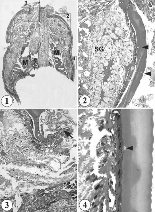
In light microscopic observations of the sections, the mass appeared to be a part of a round to elliptical-shaped organism (0.8 × 0.6 cm), and looked like a pentastomid, or an arthropod, without containing any structure of host origin (Fig. 1). It was finally identified as a species of hard tick, presumably belonging to family Ixodidae. It had a non-segmented body, well developed skeletal musculature of 160 µm in thickness in the middle part of the body (Fig. 1), well developed salivary glands in anterior half of the body (Fig. 2), and a capitulum (Fig. 3). Its external surface was covered with a thick cuticle of about 80 µm in thickness, which consisted of 5 layers, including an outermost thin layer with saw-like annulations, 3 inner layers of different thickness and structures, and the innermost layer with pore canals (Fig. 4). No legs were found sectioned.
The patient has been residing in Seoul city, Korea, since birth. The patient's past history of note is that his family took a trip to Guam Island, Indonesia, for 4 days 17 months ago. During the trip, they took a jungle river cruise in Talofofo River. Clinical examinations, including X-rays, magnetic resonance images, and blood examination, revealed no abnormalities. Other family members did not complain any symptoms similar to the boy.
DISCUSSION
For identification of arthropods including ticks, gross morphological characteristics are highly important. However, in the present case, the clinician did not have any experience with tick infestation, so simply regarded the scalp mass as a dermoid cyst and excised. We regret that the excised mass was processed for routine histological sections without gross morphological observations in the clinical laboratory. However, based on sectioned morphologies, the mass has been identified as a tick, presumably belonging to Ixodidae, i.e., hard tick.
Similar misinterpretation of tick infestations as masses of other origins is at times encountered (Pyeon and Kim, 1987). In such a case, the tick is sectioned and histological slides are made, in which case the diagnosis becomes more difficult. Nevertheless, sectional morphologies of ticks are scarcely available among the world literature. Pyeon and Kim (1987) described morphological characteristics of the cuticle layer of a hard tick based on 50 serial sections. The cuticle was composed with 5 layers, including the outermost layer characterized by saw-like annulations and the innermost basal layer having many glandular structures with basophilic granules and connected with outside through pore canals (Pyeon and Kim, 1987). In our case, the morphology of the cuticle is almost identical with the previous report of a hard tick (Pyeon and Kim, 1987). The presence of well developed musculatures and salivary glands supports the diagnosis. Moreover, in our specimens, small pieces of the capitulum were found sectioned near the anterior end of the body, and thus the capitulum is suggested to be protruded out of the anterior end of the body. By contrast, in soft ticks, the capitulum is extended from some distance beneath the anterior end of the body (Belding, 1965).
Hard ticks have a wide host range and the duration of attachment to the host skin for blood sucking is usually longer than soft ticks (Chae et al., 2000). Hence, human tick infestations are predominantly caused by hard ticks of Ixodidae. The genera of Ixodidae known to be injurious to man include Ixodes, Haemaphysalis, Amblyomma, Dermacentor, Hyalomma, Rhipicephalus, and Boophilus (Belding, 1965). In the Republic of Korea, only 2 genera, Ixodes and Haemaphysalis, were recorded from human infestations (Chae et al., 2000; Yun et al., 2001).
With regard to human tick infestations in the Republic of Korea, a total of 38 cases have been reported in the literature since 1982 (Kang et al., 1982; Lee et al., 1989; Yun et al., 2001; Hyun et al., 2002; Ko et al., 2002; Kim et al., 2003; Lee et al., 2004). Most cases were caused by Ixodes (30 cases) and Haemaphysalis (5). In the remaining 3 cases the genera of the tick were unidentified. In Ixodes, 3 species including I. nipponensis (21 cases), I. monospinosus (1), and I. persulcatus (1) were identified (species were unidentified in the remaining 7 cases), and in Haemaphysalis, H. longicornis (4) and H. flava (1) were recorded.
Yun et al. (2001) summarized 31 tick infestation cases from the first report in 1982 in terms of the predilection ages. They showed that tick infestations were more common in elderly ages over 51 years (17 of 31 cases) than younger ages. However, infestations among children younger than 10 years were not uncommon (5 out of 31 cases). We add a case of tick infestation in a 4 year-old child. With regard to the predilection site of infestation, the body trunk was the most common, which was followed by the head and the neck. In our case, the tick was found on the scalp, which is easily exposed to outsides.
The exact place of tick infestation in our case is uncertain. The patient's family had a past history of trip to Guam Island 17 months previously. Search of literature on ticks in Guam shows infestation of animals with hard tick, like Boophilus annulatus, Boophilus microplus, Rhipicephalus sanguineus, Amblyomma cyprium, and Amblyomma squamosum, and soft tick like Ornithodoros capensis (Kohl, 1953). However, no human tick infestation has been reported from Guam. It is of note that after completing their engorgement on the host skin for 7 to 12 days, most Ixodes ticks detach from the host and begin next developmental stages or oviposition (Belding, 1965; Chae et al., 2000). Thus, the possibility of tick infestation in Guam in our case is very low; the travel was 17 months previously to the notification of the tick on the scalp.
For early detection of tick infestations, careful attention should be paid, particularly in young children. During the time when ticks suck blood on the human skin, most patients hardly realize the infestation, since ticks slowly penetrate the skin with their hypostome and chelicerae, and inject saliva containing anticoagulants and anesthetic substance to the skin (Chae et al., 2000). Even after the tick is separated from the skin, careful attention is needed, because the capitulum remained in the skin could cause the tick-bite pyrexia (Chae et al., 2000). In addition, possibility of transmission of tick-borne diseases should be kept in mind, for example, Lyme disease, caused by B. burgdorferi (Lee et al., 1993; Kim et al., 1999; Lee et al., 2003; Kim and Kim, 2005), is transmitted by Ixodes tick bites (Park et al., 1992).