With regard to the intestinal histopathology of hosts infected with intestinal trematodes, villous atrophy and crypt hyperplasia, with inflammatory reactions of the villous stroma are the most important findings (Chai and Lee, 2002). Gymnophalloides seoi (Digenea: Gymnophallidae) is an intestinal trematode that infects humans in the Republic of Korea (Chai et al., 2003), and is transmitted by the oyster Crassostrea gigas (Lee et al., 1995). Histopathological changes caused by G. seoi infection are similar to those observed in other intestinal trematode infections; however, the degree of histopathology was reported to be relatively mild (Chai et al., 2001).
Since the first report of a patient with acute pancreatitis being infected with G. seoi (Lee et al., 1993), attention has been paid to the possibility of mucosal tissue invasion and extraintestinal migration by G. seoi (Chai et al., 2001, 2003). It is noteworthy that other species of gymnophallids have been recovered from the bursa Fabricii, gallbladder, and intestines, of shore birds (Yamaguti, 1971; Schell, 1985). Therefore, G. seoi has been suspected to be capable of causing infections in the pancreatic duct of man and animals. However, few studies have been performed to elucidate this point. Here, we incidentally found a human case infected with G. seoi, in which the fluke had invaded mucosal lymphoid tissue.
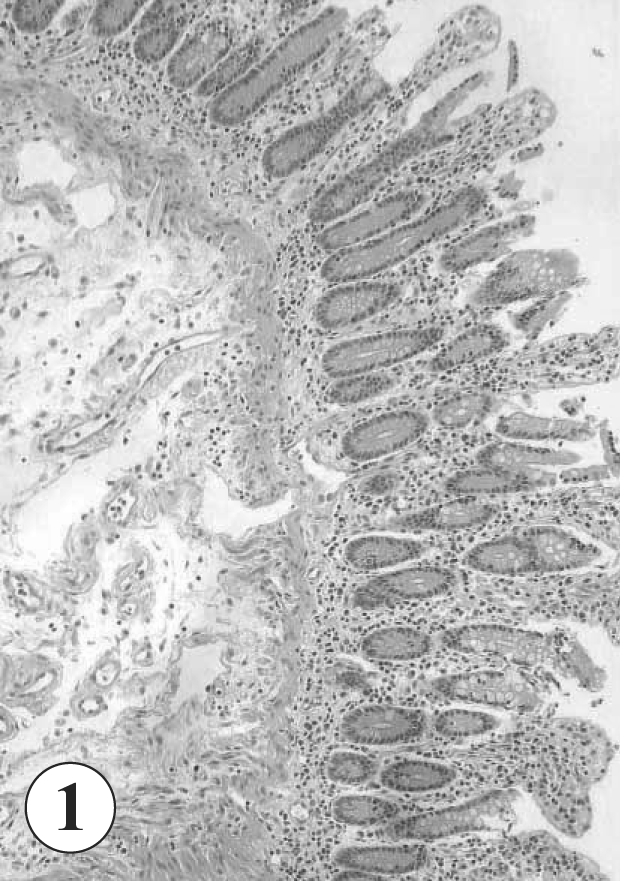
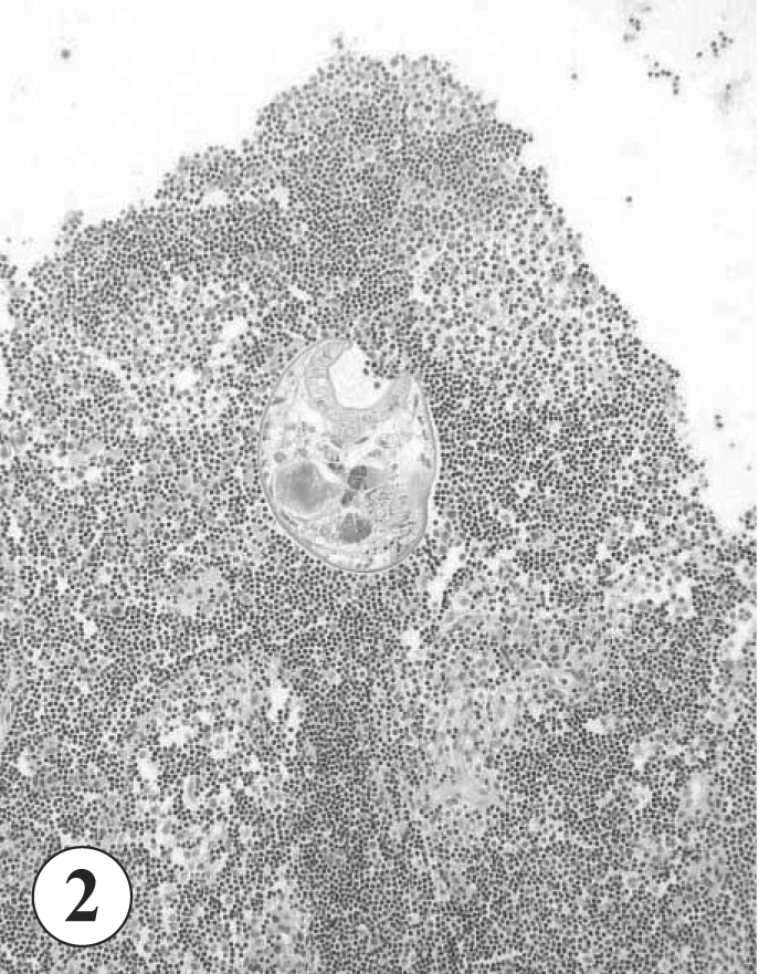
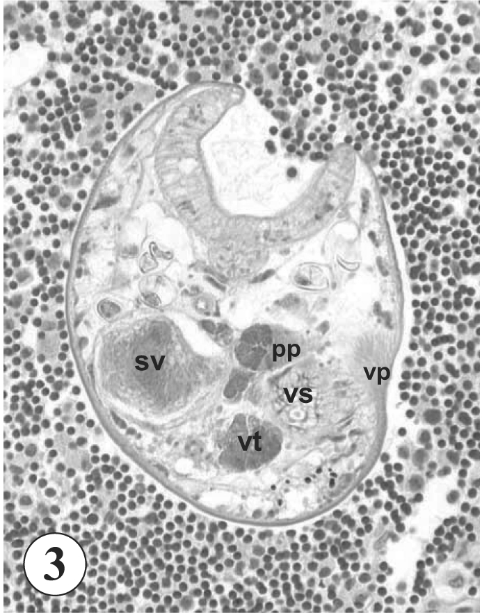
A 65-year old Korean man, living in Mokpo-city, Jeollanam-do, visited a local clinic with clinical complaints of right upper quadrant pain and indigestion in November, 2002. He also suffered from anorexia and dyspepsia. A fecal examination conducted at the time revealed no special parasite eggs or cysts. Colonoscopy and biopsy revealed the presence of a carcinoma of neuroendocrine origin at the ascending colon. He was transferred to Samsung Medical Center, where liver metastasis of the carcinoma was discovered. A palliative right hemicolectomy, small bowel resection, and anastomosis were performed, and the surgical pathology specimen revealed inflammatory reactions including villous atrophy and crypt hyperplasia of the excised colonic mucosa (Fig. 1). Moreover, an adult fluke of G. seoi was found in sectioned lymphoid tissue of the colonic wall (Figs. 2-3). The worm showed a characteristic appearance of its large oral sucker, small ventral sucker, seminal vesicle, pars prostatica, vitellaria, and a ventral pit surrounded by strong muscle fibers (Fig. 3), and was diagnosed as an adult G. seoi. The patient was treated with 10 mg/kg praziquantel, but unfortunately died due to aspiration pneumonia 3 months after the palliative operation.
Difficulty was encountered confirming the diagnosis of G. seoi infection, based on only the sectional morphology. In particular, the possibility of Parvatrema timondavidi infection should be ruled out, although no human case of P. timondavidi infection has been reported. The 2 gymnophallid species can be differentiated by the presence (G. seoi) or absence (P. timondavidi) of the ventral pit, a small inconspicuous (G. seoi) or wide (P. timondavidi) genital pore, a more posterior testes location (P. timondavidi), paired (G. seoi) or a single cluster (P. timondavidi) of vitellaria, and frequently bipartite (G. seoi) or undivided (P. timondavidi) seminal vesicle (Lee et al., 1993; Yu et al., 1993). In the worm section, the genital pore and testes were invisible, and the number of vitellaria and the shape of the seminal vesicle were unrecognizable. However, the presence of a ventral pit was suggested, by the presence of strong muscle fibers near the ventral sucker in Fig. 3. Moreover, the patient's residence, i.e., Mokpo-city, supports the diagnosis of G. seoi infection, since it is located near the highest endemic area of G. seoi in Shinan-gun (Lee et al., 1994; Chai et al., 2000). The patient is likely to have been infected with G. seoi by consuming oysters brought from Shinan-gun, Jeollanam-do (Lee et al., 1995).
In imunocompetent mice, G. seoi was observed attached to the intestinal mucosa sucking at the epithelial layer of villi with its large oral sucker (Chai et al., 2001). However, in immunocompromised hosts, some worms were found to have penetrated into the submucosa and deeper layers of the intestinal wall (Chai et al., 2001). In the present case, the patient suffered from colon cancer and received cancer chemotherapy, and therefore, he may have been, more or less, immunosuppressed. However, it is uncertain that the mucosal histopathologies, including villous atrophy and crypt hyperplasia, and symptoms complained of by this patient, i.e., right upper quadrant pain and indigestion, are the results of G. seoi infection.
It is of note that the location of the G. seoi worm found in this study was the colon rather than the small intestine. In animal hosts including mice and oystercatchers, the worms were found exclusively in the small intestines (Chai et al., 2001; Lee and Chai, 2001). In the human host, the exact habitat of G. seoi in the gastrointestinal tract was unknown. Based on the present study, it is presumed that, in humans, G. seoi may be able to infect both the small and large intestines. This is the first report of a human G. seoi infection diagnosed by a surgical pathology specimen, and demonstrates that G. seoi has the capacity to invade the human intestinal wall.