Warning: mkdir(): Permission denied in /home/virtual/lib/view_data.php on line 81
Warning: fopen(upload/ip_log/ip_log_2024-04.txt): failed to open stream: No such file or directory in /home/virtual/lib/view_data.php on line 83
Warning: fwrite() expects parameter 1 to be resource, boolean given in /home/virtual/lib/view_data.php on line 84 Identification of larval Gnathostoma obtained from imported Chinese loaches
Identification of larval Gnathostoma obtained from imported Chinese loaches
W M Sohn,*1 and S H Lee2
1Department of Parasitology, College of Medicine, Gyeongsang National University, Chinju 660-280, Korea.
Received August 16, 1996; Accepted August 26, 1996.
Abstract
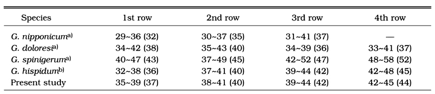
We observed the morphological characteristics and identified the species of gnathostome larvae obtained from the imported Chinese loaches. The early third-stage larvae (EL3) were collected from viscera of the loaches and a part of them were infected to mice. The advanced third-stage larvae [AdL3] were recovered from the mice at 4 weeks post-infection, both larval worms were fixed with 10% formalin, cleared in alcohol-glycerin solution, mounted with glycerin-jelly, and observed. A total of 369 EL3 were collected from viscera of 9,493 Chinese loaches. The whole body of EL3 was covered with about 190 transverse rows of minute cuticular spines and 0.624 × 0.101 mm in average size. A pair of lips were protruded at the anterior end, and the muscular esophagus and brownish intestine were followed. The characteristic head bulb was provided with 4 rows of hooklets. The average number of hooklets in the respective row was 36.7, 39.5, 41.6 and 44.3 posteriorly. AdL3 was 2.660 × 0.346 mm in average size, and retained the esophagus (about 0.755 mm length) and cervical sac (about 0.355 mm length). The average number of hooklets in the respective row on the head bulb was 39.0, 41.9, 43.9 and 45.6, posteriorly. On the basis of the morphological characteristics, they were identified as the third-stage larvae of Gnathostoma hispidum.
Figures
Figs. 1-3 Fig. 1. The early third-stage larva (EL3) of G. hispidum collected in the viscera of a loach imported from China. A: anus; CS: cervical sac; E: esophagus; HB; head bulb; I: lip. Fig. 2. The schematic drowing of EL3 of which transverse striations of minute cuticular spines were omitted. Fig. 3. The head bulb of an EL3 which retained 4 rows of hooklets with sharp-pointed end.
Figs. 4-8 Fig. 4. The advanced third-stage larva (AdL3) of G. hispidum encysted in the liver of an experimental mouse. Fig. 5. The AdL3 of G. hispidum recovered from the muscle of a mouse experimentally infected with EL3. Fig. 6. The schematic drowing of AdL3. Fig. 7. The head bulb of an AdL3. bearing 4 transverse rows of hooklets. Fig. 8. The head bulb of an AdL3. Note the 5th row of hooklets (arrow marks).
Tables
Table 1 Infection status of imported Chinese loaches with larval gnathostome
Table 2 Measurementsa) of the larval gnathostome from the Chinese loaches and comparison with those of previous authors
Table 3 Measurementsa) of the advanced third-stage larvae (AdL3) from the experimentally infected mice and comparison with those of previous authors
Table 4 Comparison of the number of hooklets on head bulbs in several species of larval Gnathostoma
References
1.
Akahane H, et al. Jpn J Parasitol 1982;31:507–516.
2.
Akahane H, et al. Jpn J Parasitol 1984;33:509–513.
3.
Ando K, Tanaka H, Taniguchi Y, Shimizu M, Kondo K. Two human cases of gnathostomiasis and discovery of a second intermediate host of Gnathostoma nipponicum in Japan. J Parasitol 1988;74(4):623–627.
4.
Araki T. Kansen Ensyou Meneki 1986;16:110–111.
6.
Chen HT. Lingnan Sci J 1936;15:31–44.
7.
Daengsvang S. Further observations on the experimental transmission of Gnathostoma spinigerum. Ann Trop Med Parasitol 1968;62(1):88–94.
9.
Demitsu T, et al. Rinshohifuka 1985;39:255–260.
10.
Huang WC, et al. Jpn J Parasitol 1986;35(3):223–227.
11.
Kim CH. [The Infection Status Of Sparganum And Gnathostoma In Frogs Of Southern Part Of Korea]. Korean J Parasitol 1983;21(1):83–86.
12.
Kim YK. Bull Pusan Nat Univ 1973;15:111–116.
13.
Koga M, et al. Jpn J Parasitol 1985;34:361–370.
14.
Lee SH, Hong ST, Chai JY. Korean J Parasitol 1988;26(1):33–38.
16.
Miyazaki I. On the genus Gnathostoma and human gnathostomiasis, with special reference to Japan. Exp Parasitol 1960;9:338–370.
18.
Miyazaki I, et al. Acta Med (Fukuoka) 1952;22:467–473.
19.
Morita H, et al. J Nara Med Assoc 1984;35:607–619.
20.
Nawa Y, Imai J, Ogata K, Otsuka K. The first record of a confirmed human case of Gnathostoma doloresi infection. J Parasitol 1989;75(1):166–169.
21.
Sohn WM, Kho WG, Lee SH. Larval Gnathostoma nipponicum found in the imported Chinese loaches. Korean J Parasitol 1993;31(4):347–352.
22.
Takakura Y, et al. Jpn J Parasitol 1985;34(4):211–218.