Warning: mkdir(): Permission denied in /home/virtual/lib/view_data.php on line 81

Warning: fopen(upload/ip_log/ip_log_2024-04.txt): failed to open stream: No such file or directory in /home/virtual/lib/view_data.php on line 83

Warning: fwrite() expects parameter 1 to be resource, boolean given in /home/virtual/lib/view_data.php on line 84
A Survey of Intestinal Protozoan Infections among Gastroenteritis Patients during a 3-Year Period (2004-2006) in Gyeonggi-do (Province), South Korea
| Home | E-Submission | Sitemap | Contact us |  
top_img
Korean J Parasito Search

CLOSE

Korean J Parasito > Volume 47(3):2009 > Article
Huh, Moon, and Lim: A Survey of Intestinal Protozoan Infections among Gastroenteritis Patients during a 3-Year Period (2004-2006) in Gyeonggi-do (Province), South Korea

Abstract

The incidence and etiology of parasite-associated gastroenteritis during 2004-2006 in Gyeonggi-do (province), South Korea was determined by means of antigen detection ELISA on 6,071 stool specimens collected from 6 general hospitals. At least 1 parasitic agent was detected in 3.4% (208/6,071) of the stool samples. Among these, Giardia lamblia was the most numerous (152 cases; 2.5%), followed by Entamoeba histolytica (25 cases; 0.4%), Cryptosporidium parvum (23 cases; 0.4%), and mixed infections (8 cases; 0.1%). Patients aged 1-5 years had the largest proportion (69.2%; 144/208) of parasite-positive stool specimens. Parasite-mediated gastroenteritis was most common from June to September. The detection rate gradually increased from 2004 to 2006. This study shows that parasite-mediated gastroenteritis may be significant among children in Korea and that parasite infection surveillance should be constantly performed.

Acute diarrhea is one of the most common childhood illnesses in both developing and developed countries. The etiologic agents of diarrhea include viruses, bacteria, and parasites [1]. Among them, viral infection is the most common cause, followed by bacterial and parasitic infections [2]. Giardia lamblia, Entamoeba histolytica, and Cryptosporidium parvum are the major parasitic agents [1]. Although they are not serious life-threatening parasites, they are still important infectious agents, especially to infants. Most survey reports on gastroenteritis outbreaks in South Korea have mainly focused on viral and bacterial infections [3-6], while gastroenteritis caused by intestinal protozoan parasites has not been well studied. We describe here the distribution of intestinal protozoan parasites causing gastroenteritis and variations with patient age and seasons.
This epidemiologic survey was performed for 3 years in Gyeonggi-do (province) of South Korea. In this province, a region located around the capital city of Korea, approximately 11 million people, 25% of the country's population, lives. A total of 6,071 stool specimens were collected from patients with gastroenteritis in 6 general hospitals in Gyeonggi-do during 2004-2006. Cysts of G. lamblia and E. histolytica, and oocyusts of C. parvum were detected by antigen detection ELISA specific for Cryptosporidium, Giardia, and E. histolytica/dispar (IVD Research, Carlsbad, California, USA).
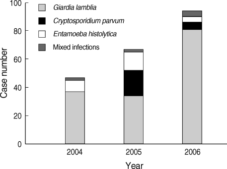
Of the 6,071 patients, 54.7% were below 5 years of age. The intestinal protozoans were detected in 208 (3.4%) samples. The detection rate increased annually, from 1.7% (47/2,739) in 2004 to 4.0% (67/1,675) in 2005 and to 5.7% (94/1,657) in 2006. One of the reasons might be the improvement of diagnostic skills each year. The most predominant parasite species was G. lamblia (152 cases; 2.5%), followed by Entamoeba histolytica (25 cases; 0.4%), Cryptosporidium parvum (23 cases; 0.4%), and mixed infections (8 cases; 0.1%). Mixed infections represented Giardia/Entamoeba, Cryptosporidium/Giardia, and Cryptosporidium/Giardia/Entamoeba. The yearly distributions showed that G. lamblia was the most prevalent species throughout the 3 years (Fig. 1).
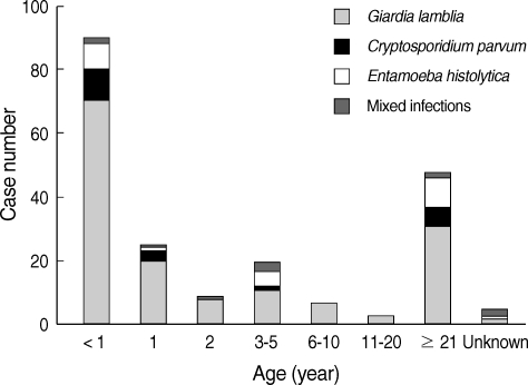
The distribution of parasitic agents according to age is shown in Fig. 2. Of the total number of detected samples, 23.1% (48 of 208) were obtained from adults aged 21 years of age and older, while 69.2% (144 of 208) were obtained from children 5 years of age and younger. Of the latter, 43.3% (90 of 208) of the positives were obtained from infants less than 1-year-old. The majority of mixed infections were found in under 5-year-old children. All parasitic co-infections were infected with G. lamblia.
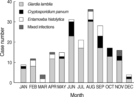
The incidence in winter and spring months (December-May) and summer and autumn months (June-November) were 28.4% and 71.6%, respectively (Fig. 3). The total prevalence in summer-autumn was 2.5 times higher than that in winter-spring. G. lamblia was detected throughout the year, with detection rate being higher in summer-autumn, while E. histolytica and C. parvum were rarely detected in winter (December-May). Bacterial agents have been detected frequently in summer and autumn like parasitic infections, whereas viral gastroenteritis is prevalent in the winter-spring season [6,7].
Presently, parasitic infections causing gastroenteritis were a special concern to children aged less than 5 years. This is consistent in many studies which have shown that children under 5 years of age are the most vulnerable to parasitic gastroenteritis worldwide [8,9]. In our study, G. lamblia was the most prevalent etiologic agent, consistent with the worldwide trend [8]. We regret that morphological identification of G. lamblia and E. histolytica cysts, and of C. parvum oocysts, was not performed in positive samples. However, our results may indicate usefulness and feasibility of the antigen detection ELISA for surveys of intestinal protozoan infections using fecal samples.
We also detected co-infections with parasites and viruses. The viral agents included rotavirus, adenovirus, astrovirus, and norovirus, which are the major viral etiologic agents of gastroenteritis. The viruses were detected using PCR for adenovirus and reverse transcription-PCR for astrovirus, rotavirus, and norovirus [10-15]. The co-infections rates with viruses and G. lamblia, E. histolytica, C. parvum, or mixed infections were 27.6% (42/152), 32.0% (8/25), 47.8% (11/23), and 75.0% (6/8), respectively. Among 6,071 stool specimens, at least 1 viral agent was detected in 30.0% (1,819/6,071) of the stool samples. Among these, rotavirus was the most numerous (53.5%), followed by norovirus (12.6%), astrovirus (7.3%), and enteric adenovirus (0.3%) [16]. The etiologic agents for diarrhea of the rest (66.6% of stool specimens) were unknown.
The present study was a comprehensive longer-term (3 years) investigation of etiologic agents causing parasitic gastroenteritis in South Korea. While useful, the full impact of the study was blunted by the manner of specimen collection. Although stool specimens were collected from the patients with gastroenteritis, there might be some unidentified cases. Thus, our prevalence and incidence numbers might be lower than the total cases of parasitic gastroenteritis in the community. Parasitic contamination in water and foods may escape detection because the food standard regulations are usually limited to bacterial and viral contaminations. Nowadays many foods are imported from foreign locations globally. Hence, foreign-derived parasitic agents might be circulating in the market. Therefore, surveillance of parasitic pathogens should be constantly performed to monitor circulating parasitic agents in the community.

REFERENCES

1. Aranda-Michel J, Giannella RA. Acute diarrhea: a practical review. Am J Med 1999;106:670-676. PMID: 10378626.
crossref pmid
2. Centers for Disease Control and Prevention, Republic of Korea. EnterNet-Korea Surveillance Report 2007;12.

3. Song MO, Kim KJ, Chung SI, Lim I, Kang SY, An CN, Kim W. Distribution of human group a rotavirus VP7 and VP4 types circulating in Seoul, Korea between 1998 and 2000. J Med Virol 2003;70:324-328. PMID: 12696125.
crossref pmid
4. Kang JO, Kilgore P, Kim JS, Nyambat B, Kim J, Suh HS, Yoon Y, Jang S, Chang C, Choi S, Kim MN, Gentsch J, Bresee J, Glass R. Molecular epidemiological profile of rotavirus in South Korea, July 2002 through June 2003: emergence of G4P[6] and G9P[8] strains. J Infect Dis 2005;192:S57-S63. PMID: 16088806.

5. Cho SH, Shin HH, Choi YH, Park MS, Lee BK. Enteric bacteria isolated from acute diarrheal patients in the Republic of Korea between the year 2004 and 2006. J Microbiol 2008;46:325-330. PMID: 18604503.
crossref pmid
6. Cho SH, Kim JH, Kim JC, Shin HH, Kang YH, Lee BK. Surveillance of bacterial pathogens associated with acute diarrheal disease in the Republic of Korea during one year, 2003. J Microbiol 2006;44:327-335. PMID: 16820763.

7. Seo JK, Sim JG. Overview of rotavirus infections in Korea. Pediatr Intl 2000;42:406-410.
crossref pmid
8. Schuster H, Chiodini PL. Parasitic infections of the intestine. Curr Opin Infect Dis 2001;14:587-591. PMID: 11964880.
crossref pmid
9. Lengerich EJ, Addiss DG, Juranek DD. Severe giardiasis in the United States. Clin Infect Dis 1994;18:760-763. PMID: 8075266.
crossref pmid
10. Belliot G, Laveran H, Monroe SS. Detection and genetic differentiation of human astroviruses: phylogenetic grouping varies by coding region. Arch Virol 1997;142:1323-1334. PMID: 9267446.
crossref pmid
11. Kojima S, Kageyama T, Fukushi S, Hoshino FB, Shinohara M, Uchida K, Natori K, Takeda N, Katayama K. Genogroup-specific PCR primers for detection of Norwalk-like viruses. J Virol Methods 2002;100:107-114. PMID: 11742657.
crossref pmid
12. Pring-Akerblom P, Adrian T. The hexon genes of adenoviruses of subgenus C: comparison of the variable regions. Res Virol 1993;144:117-127. PMID: 8511397.
crossref
13. Pring-Akerblom P, Adrian T, Köstler T. PCR-based detection and typing of human adenoviruses in clinical samples. Res Virol 1997;148:225-231. PMID: 9201813.
crossref
14. Gentsch JR, Glass RI, Woods P, Gouvea V, Gorziglia M, Flores J, Das BK, Bhan MK. Identification of group A rotavirus gene 4 types by polymerase chain reaction. J Clin Microbiol 1992;30:1365-1373. PMID: 1320625.
pmid pmc
15. Gouvea V, Glass RI, Woods P, Taniguchi K, Clark HF, Forrester B, Fang ZY. Polymerase chain reaction amplification and typing of rotavirus nucleic acid from stool specimens. J Clin Microbiol 1990;28:276-282. PMID: 2155916.
pmid pmc
16. Huh JW, Kim WH, Moon SG, Lee JB, Lim YH. Viral etiology and incidence associated with acute gastroenteritis in a 5-year survey in Gyeonggi province, South Korea. J Clin Virol 2009;44:152-156. PMID: 19131274.
crossref pmid
Fig. 1
Yearly distribution of predominant parasitic agents causing gastroenteritis during 2004-2006.
kjp-47-303-g001.jpg
Fig. 2
Distribution of parasitic agents of gastroenteritis by age in Gyeonggi-do (province), South Korea, during 2004-2006. Mixed infections represent Giardia/Entamoeba, Cryptosporidium/Giardia, and Cryptosporidium/Giardia/Entamoeba.
kjp-47-303-g002.jpg
Fig. 3
Distribution of parasitic agents of gastroenteritis by month in Gyeonggi-do (province), South Korea, during 2004-2006. Mixed infections represent Giardia/Entamoeba, Cryptosporidium/Giardia, and Cryptosporidium/Giardia/Entamoeba.
kjp-47-303-g003.jpg
TOOLS
PDF Links  PDF Links
PubReader  PubReader
ePub Link  ePub Link
XML Download  XML Download
Full text via DOI  Full text via DOI
Download Citation  Download Citation
  Print
Share:      
METRICS
14
Crossref
14
Scopus
7,516
View
118
Download
Editorial Office
Department of Molecular Parasitology, Samsung Medical Center, School of Medicine, Sungkyunkwan University,
2066 Seobu-ro, Jangan-gu, Suwon 16419, Gyeonggi-do, Korea.
Tel: +82-31-299-6251   FAX: +82-1-299-6269   E-mail: kjp.editor@gmail.com
About |  Browse Articles |  Current Issue |  For Authors and Reviewers
Copyright © 2024 by The Korean Society for Parasitology and Tropical Medicine.     Developed in M2PI