Warning: mkdir(): Permission denied in /home/virtual/lib/view_data.php on line 81
Warning: fopen(upload/ip_log/ip_log_2024-04.txt): failed to open stream: No such file or directory in /home/virtual/lib/view_data.php on line 83
Warning: fwrite() expects parameter 1 to be resource, boolean given in /home/virtual/lib/view_data.php on line 84 Antigenic localities in the tissues of the young adult worm of Paragonimus westermani using immunogold labeling method
Antigenic localities in the tissues of the young adult worm of Paragonimus westermani using immunogold labeling method
O S Kwon,J S Lee,H J Rim and S J Kim*
Department of Parasitology and Institute for Tropical Endemic Diseases, College of Medicine, Korea University, Seoul 136-701, Korea.
Abstract
In order to observe the antigenic localization in the tissues of the young adult Paragonimus westermani, immunogold labeling method was applied using serum immunoglobulins(IgG) of the dog which infected with isolated metacercariae from Cambaroides similis. The sectioned worm tissue was embedded in Lowicryl HM 20 medium and stained with infected serum IgG and protein A gold complex (particle size; 12 nm). It was observed by electron microscopy at each tissues of the worm. The gold particles were not observed on the basal lamina of the tegument, interstitial matrix of the parenchyma, the muscle tissue and mitochondria of the tegument. The gold particles were specifically labeled in the secretory granules in the vitelline cells. They were predominantly labeling on the epithelial lamela and lumen of caecum. The above finding showed that antigenic materials in young adult worm tissue were specifically concentrated on the tegumental syncytium as well as cytoplasm of tegumental cells.
Figures
Figs. 1-2 Fig. 1. Electron micrograph of the tegument of a worm which was reacted with dog IgG from noninfected control showed the tegumental syncytium(TS), basal layer(BL), circular muscle (CM) layer, longitudinal muscle(LM) layer, interstitial matrix(IM) and tegumental cell cytoplasm (TCC). Gold particles were not labeled on the tegument or other portions of the tissue. Bar = 1 µm (×17,000)
Fig. 2. The tegumental tissue of the worm reacted with specific antibody(IgG) from infected dog. Gold particles were specifically labeled in the tegumental syncytium and cytoplasm of the tegumental cell. Bar = 1 µm(×28,000)
Figs. 3-4 Fig. 3. The tegumental cell of the tegument reacted with control group dog IgG from infected control. Gold particle was not labeled on the cytoplasm of the cell. Bar = 1 µm(×17,000)
Fig. 4. The tegumental cell of the tegument reacted with specific antibody(IgG) from infected dog. Specific label was present in the cytoplasm of the tegumental cell. Bar=1 µm(×17,000)
Figs. 5-6 Fig. 5. The vitelline gland of a worm which was reacted with control group dog IgG from noninfected control. Gold particles were not labeled on the cytoplasm and secretory granules of vitelline gland cell. Bar=1 µm(×28,000)
Fig. 6. The vitelline gland of a worm which was reacted with specific antibody(IgG) from infected dog. Gold particles were very specifically labeled on the secretory granules of vitelline gland cell cytoplasm. Bar=1 µm(×28,000)
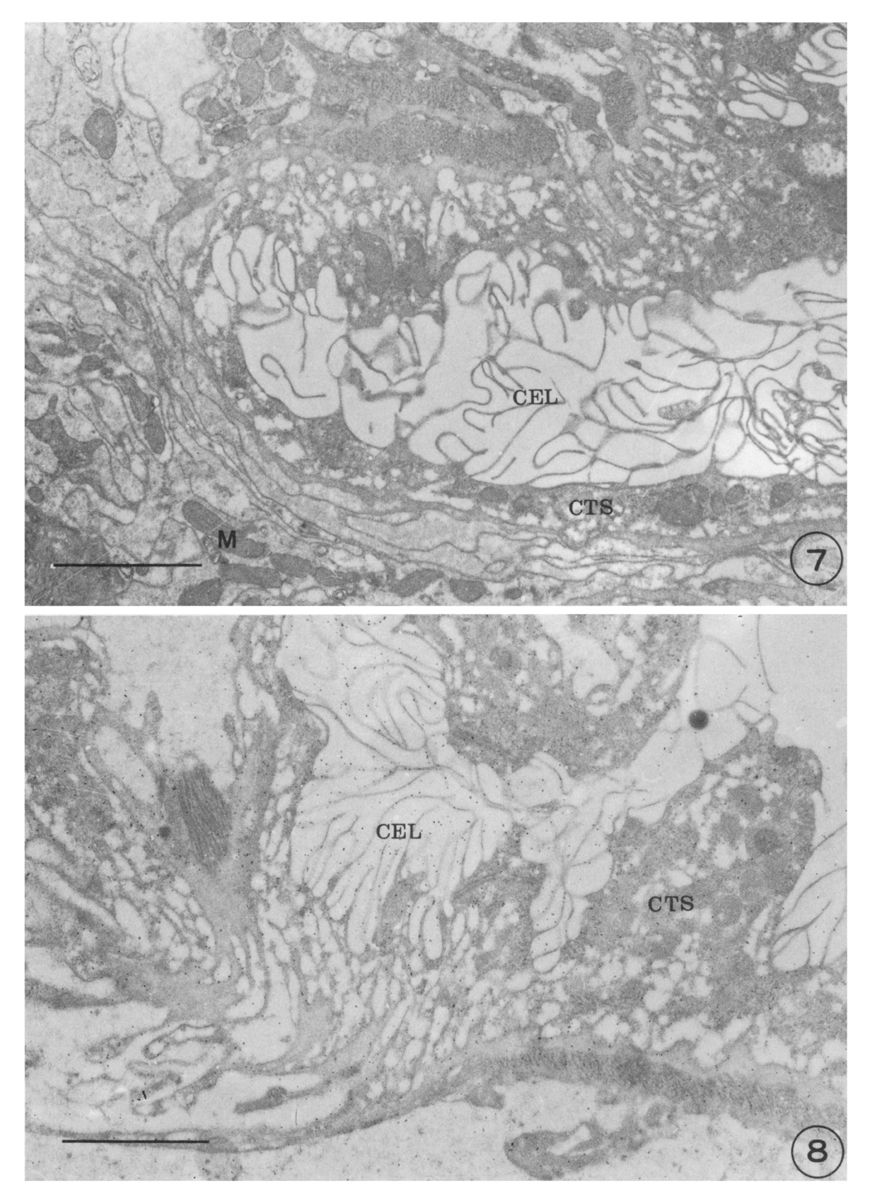
Figs. 7-8 Fig. 7. The caecal section of a worm reacted with dog IgG from noninfected control. Gold particles were not labeled on the all area of the caecum. Bar=1 µm(×17,000)
Fig. 8. The caecal section of a worm reacted with specific antibody(IgG) from infected dog. Gold particles were particles were predominantly labeled on the caecum tegument syncytium. Bar=1 µm(×17,000)
Tables
Table 1 Quantitative density of the labeled gold particles in the tissues of P. westermani reacted with antibody (IgG)* obtained from dogs infected with P. westermani
References
1.
Cho KM, et al. Yonsei Rep Trop Med 1976;7:26–37.
2.
Cho SY, Hong ST, Rho YH, Choi SY, Han YC. Application of micro-ELISA in serodiagnosis of Human paragonimiasis. Korean J Parasitol 1981;19(2):151–156.
3.
Choi WY, Lee WK, Lee OR. [Indirect Fluorescent Antibody Test For Diagnosis Of Paragonimiasis]. Korean J Parasitol 1975;13(2):152–158.
4.
Choi WY, Lee OR. [Agar-Gel Precipitin Reactions In Experimental Paragonimiasis]. Korean J Parasitol 1981;19(2):101–108.
5.
Choi WY, Yoo JE, Nam HW, Choi HR. [Purification of antigenic proteins of Paragonimus westermani and their applicability to experimental cat paragonimiasis]. Korean J Parasitol 1986;24(2):177–186.
6.
de Water R, Fransen JA, Deelder AM. Ultrastructural localization of the circulating anodic antigen in the digestive tract of Schistosoma mansoni using monoclonal antibodies in an immunogold labeling procedure. Am J Trop Med Hyg 1986;35(3):549–558.
7.
Fujino T, et al. Jpn J Parasitol 1989;38(5):263–270.
8.
Fukuda K, Fujino T, Hamajima F. Crystalline inclusions in the subtegumental cells of the adult lung fluke, Paragonimus westermani. Z Parasitenkd 1982;68(2):235–238.
9.
Huer B, Kim SI, Kang SY, Cho SY. Electrophoretic patterns of proteins from Paragonimus westermani in early developmental stages. Korean J Parasitol 1985;23(2):189–196.
10.
Imai J. Trop Med 1979;21(2):45–55.
11.
Chu BD, Rim HJ, Kim SJ. [A study on the body fluid antigen of Clonorchis sinensis using immunogold labeling method]. Korean J Parasitol 1990;28(1):11–23.
12.
Joo KH, Ahn H, Chung MS, Rim HJ. Demonstration of species-specific and cross reactive components of Paragonimus westermani crude worm antigen by EITB. Korean J Parasitol 1989;27(1):9–14.
13.
Kim DC, et al. Rep NIH Korea 1982;19:109–114.
14.
Kim SI, Ko EK, Kang SY, Cho SY. [Antigenicity of the soluble egg antigen of Paragonimus westermani]. Korean J Parasitol 1986;24(1):49–54.
15.
Kim SJ, et al. Korean J Zool 1988;31:62–70.
16.
Kim SJ, Lee KO, Takamiya S, Capaldi RA. Mitochondrial myopathy involving ubiquinol-cytochrome c oxidoreductase (complex III) identified by immunoelectron microscopy. Biochim Biophys Acta 1987;894(2):270–276.
17.
Lee OR, Chang JK. [ELISA of paragonimiasis in cat by crude and purified antigens of Paragonimus westermani]. Korean J Parasitol 1986;24(2):187–193.
18.
Lee SH, Sung SH, Chai JY. [Immunohistochemical study on the antigenicity of body compartments of Paragonimus westermani]. Korean J Parasitol 1989;27(2):109–117.
19.
von Lichtenberg F, Bawden MP, Shealey SH. Origin of circulating antigen from the schistosome gut. An immunofluorescent study. Am J Trop Med Hyg 1974;23(6):1088–1091.
20.
Nash TE. Localization of the circulating antigen within the gut of Schistosoma mansoni. Am J Trop Med Hyg 1974;23(6):1085–1087.
21.
Roth J. The preparation of protein A-gold complexes with 3 nm and 15nm gold particles and their use in labelling multiple antigens on ultra-thin sections. Histochem J 1982;14(5):791–801.
22.
Roth J. Applications of immunocolloids in light microscopy. Preparation of protein A-silver and protein A-gold complexes and their application for localization of single and multiple antigens in paraffin sections. J Histochem Cytochem 1982;30(7):691–696.
23.
Sugiyama H, Sugimoto M, Akasaka K, Horiuchi T, Tomimura T, Kozaki S. Characterization and localization of Paragonimus westermani antigen stimulating antibody formation in both the infected cat and rat. J Parasitol 1987;73(2):363–367.
24.
Sun T, et al. Jap J Med Sci Biol 1969;22:263–272.
25.
Yogore MG, et al. Am J Trop Med Hyg 1965;14(4):586–591.
26.
Yokogawa M, et al. Jpn J Parasitol 1962;11(2):117–122.