Warning: mkdir(): Permission denied in /home/virtual/lib/view_data.php on line 81
Warning: fopen(upload/ip_log/ip_log_2024-04.txt): failed to open stream: No such file or directory in /home/virtual/lib/view_data.php on line 83
Warning: fwrite() expects parameter 1 to be resource, boolean given in /home/virtual/lib/view_data.php on line 84 Experimental life history of Spirometra erinacei
Department of Parasitology and Institute of Endemic Diseases, Seoul National University College of Medicine, Seoul 110-460, Korea.
**Present address: Department of Parasitology, College of Medicine, Inje University, Pusan 614-735, Korea.
Abstract
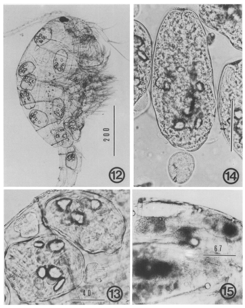
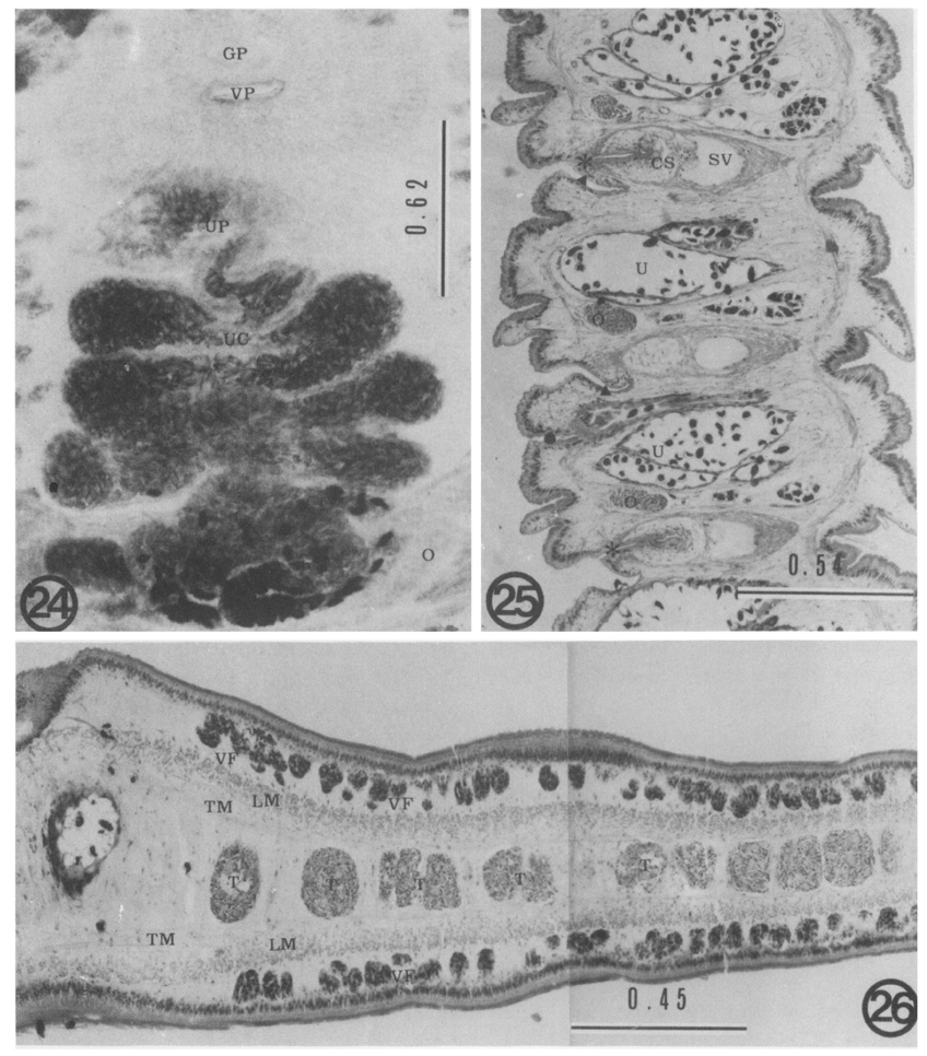
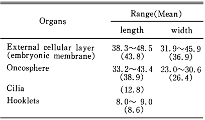
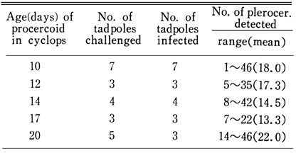
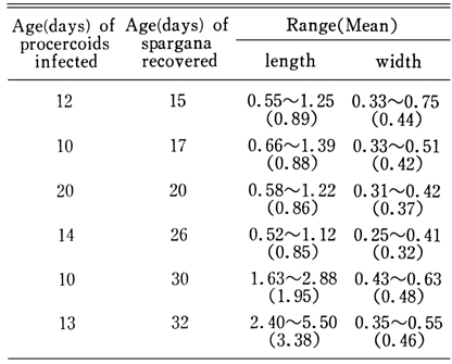
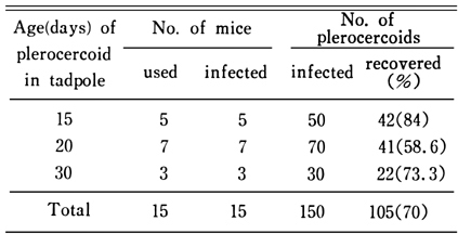
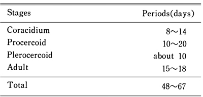
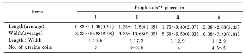
The complete life cycle of Spirometra erinacei has been experimentally maintained in the laboratory. The cyclops were reared as the first intermediate host, and the tadpoles of Rana nigromaculata as the second intermediate host. ICR mice were used as another second host. The experimental definitive hosts were dogs and cats. Maturation and hatching of the eggs took 8 to 14 days by incubation at 29℃. The coracidium measured 43.8 × 36.9 µm. Mesocyclops leuckarti and Eucyclops serrulatus were susceptible to the coracidial infection. The procercoids older than 5 days in the cyclops had minute spines at the anterior end, calcium corpuscles in the body parenchyme and the cercomer at the posterior end. Procercoids 10 to 20 days old were infective to tadpoles, and 15 or 21 day old worms could infect the mice. The plerocercoids from the tadpoles at 15 days after experimental infection were pear-shaped and shorter than 1 mm in the length and were infective to mice. Fifteen to 18 days after experimental inoculation of plerocercoids to dogs or cats, the adult worms began to produce eggs. One life cycle from egg to egg needed 48 to 67 days in the laboratory. The morphology of larval or adult worms was compatible with the description of Spirometra erinacei.
Table 2 Measurenments of S. erinacei procercoids* according to the age of infection
Table 3 The infectivity of S. erinacei procercoids to tadpoles
Table 4 Measurements of spargana* collected from experimental tadpoles
Table 5 The infectivity of S. erinacei proceroids to mice
Table 6 The infectivity of S. erinacei plerocercoids to mice
Table 7 The periods for development of S. erinacei by stages
Table 8 Measurements* of S. erinacei mature proglottids from the experimentally infected dog
References
1.
Cho SY, Bae JH, Seo BS. Some Aspects Of Human Sparganosis In Korea. Korean J Parasitol 1975;13(1):60–77.
2.
Faust EC, et al. Am J Hyg 1929;9:560–583.
3.
Hong ST, Kim KJ, Huh S, Lee YS, Chai JY, Lee SH, Lee YS. The changes of histopathology and serum anti-sparganum IgG in experimental sparganosis of mice. Korean J Parasitol 1989;27(4):261–269.
4.
Iwata S. Progress of Med Parasit 1972;4:536–590.
5.
Iwata S, et al. Jpn J Parasitol 1967;16:568.
6.
Kim CH. [The Infection Status Of Sparganum And Gnathostoma In Frogs Of Southern Part Of Korea]. Korean J Parasitol 1983;21(1):83–86.
7.
Lee SH, Chai JY, Seo BS, Cho SY. Two cases of human infection by adult of Spirometra erinacei. Korean J Parasitol 1984;22(1):66–71.
8.
Li CH. Am J Hyg 1929;10(3):527–550.
9.
Mueller JF. Am J Trop Med 1938;18:41–66.
10.
Suzuki N, et al. Jpn J Parasitol 1982;31(1):23–26.
11.
Weinstein PP, Krawczyk HJ, Peers JH. Sparganosis in Korea. Am J Trop Med Hyg 1954;3(1):112–129.