Warning: mkdir(): Permission denied in /home/virtual/lib/view_data.php on line 81
Warning: fopen(upload/ip_log/ip_log_2024-04.txt): failed to open stream: No such file or directory in /home/virtual/lib/view_data.php on line 83
Warning: fwrite() expects parameter 1 to be resource, boolean given in /home/virtual/lib/view_data.php on line 84 Tegumental ultrastructures of Paragonimus iloktsuenensis according to the developmental stages
Tegumental ultrastructures of Paragonimus iloktsuenensis according to the developmental stages
S H Lee,S J Kim,J Y Chai and W M Sohn*
Department of Parasitology and Institute of Endemic Diseases, College of Medicine,Seoul National University, Seoul 110-460, Korea.
Department of Parasitology*, College of Medicine, Inje University, Pusan 614-112, Korea.
Abstract
A scanning electron microscopic study was performed to observe the tegumental ultrastructures of Paragonimus iloktsuenensis according to its developmental stages. The metacercariae were obtained from the liver of the brackish water crab, Sesarma dehaani. Juvenile and adult P. iloktsuenensis were recovered from the experimental rats on 2, 4 and 8 weeks after infection. The findings were summarized as follows: 1. The excysted metacercariae were characteristically gourd-shape, with their whole body surface beset with numerous spade-shape spines. The large, type II sensory papillae (non-ciliated round swellings) were arranged along the rim of the oral and ventral suckers, 11-12 and 6-8 in numbers respectively. 2. Two-week old juvenile worms, recovered chiefly from the liver of the experimental rats, were slender in body shape, with their ventral sucker near the anterior one-third level. The distribution of tegumental spines was less dense than in the excysted metacercariae. The spines were with 1-2 pointed tips and 3-4 longitudinal splits. Numerous ciliated knob-like, type I papillae were observed in both sides of the oral sucker, and 6 large, type II papillae were arranged along the rim of the ventral sucker. 3. Four-week old worms, recovered from the thoracic cavity and/or lung parenchyme of the experimental rats, were thicker than wide in body configuration, and their ventral sucker was located near the anterior one-fourth level. The tegumental spines at ventral surface were grouped, each group with 3-5 aggregated ones. The type I and type II papillae (small-sized) were distributed chiefly around the rim of two suckers. 4. Adult (eight-week old) worms, recovered from the capsules in the lung parenchyme, were very stout, and covered densely with bearfoot-like spines. At dorsal surface, cobblestone-like cytoplasmic processes were well-developed, with many tegumental spines embedded in them. It was observed in this study that the tegument of P. iloktsuenesis continued to change and differentiate as the worms grew to be adults.
Figures
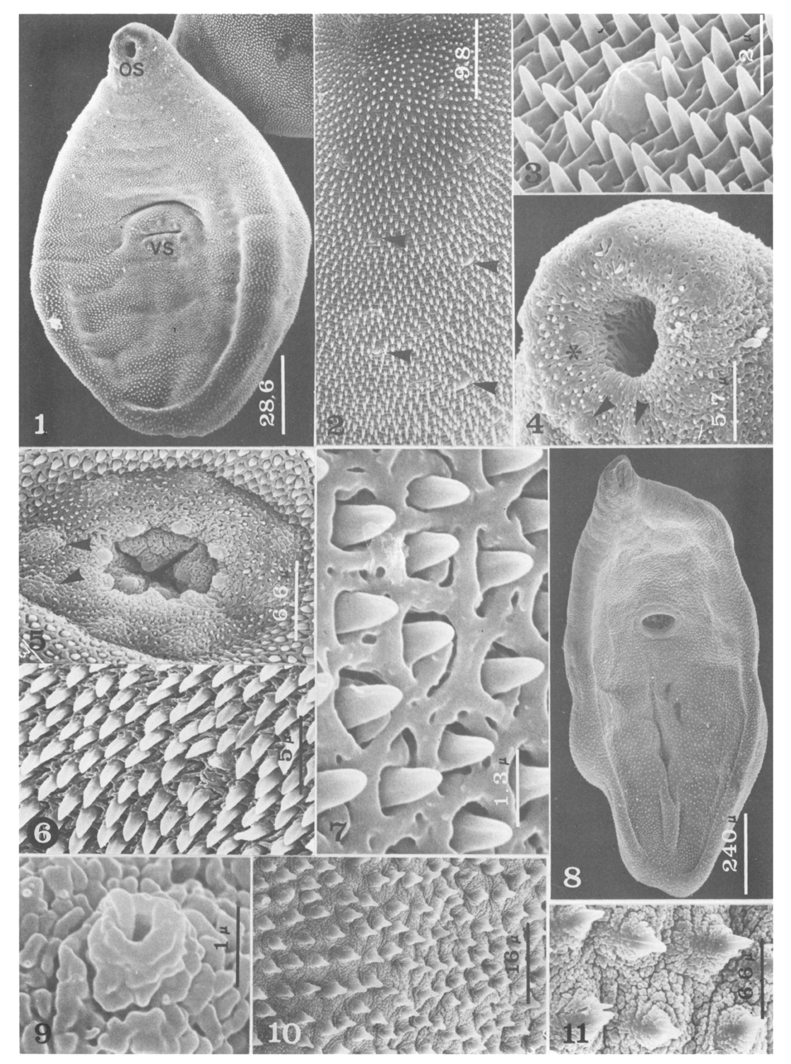
Figs. 1-11 Fig. 1. Ventral view of an excysted metacercaria of P. iloktsuenensis. OS; oral sucker, VS; ventral sucker (×700).
Fig. 2. Just behind and slightly lateral portion of the oral sucker of the excysted larva. Two rows of small, type II papillae(arrow head) are characteristically seen (×2,000).
Fig. 3. Magnification of Fig. 2. (×10,000).
Fig. 4. Oral sucker of the excysted larva having large, type II(arrow head) and pit-type papillae(asterisk) (×3,500).
Fig. 5. Ventral sucker of the excysted larva having 8 large, type II papillae(arrow head) and 8 small, type I papillae in a concentric circle (×3,000).
Fig. 6. Tegumental spines at dorso-anterior portion of the excysted larva (×4,000).
Fig. 7. Tegumental spines at dorso-posterior portion of the excysted larva (×15,000).
Fig. 8. Ventral view of a 2-week old worm (×800).
Fig. 9. Type I sensory papillae on the oral sucker of the 2-week old worm (×20,000).
Fig. 10. Tegumental spines between the oral and ventral suckers (×1,200).
Fig. 11. Magnification of Fig. 10 (×3,000). (Bar; µm)
Figs. 12-23 Fig. 12. Ventral sucker of a 2-week old worm (×1,400).
Fig. 13. Tegumental spines at posterior portion of the ventral sucker of the 2-week old worm (×4,300).
Fig. 14. Dorso-anterior tegument of the 2-week old worm (×6,000).
Fig. 15. Dorso-posterior tegument of the 2-week old worm (×1,800).
Fig. 16. Ventral view of a 4-week old worm (×400).
Fig. 17. Tegumental spines between the oral and ventral sucker of the 4-week old worm (×2,000).
Fig. 18. Magnification of Fig. 17 (×5,000).
Fig. 19. Ventral sucker of the 4-week old worm (×430).
Fig. 20. Tegumental spines at ventro-posterior portion of the 4-week old worm (×260).
Fig. 21. Magnification of Fig. 20 (×3,000).
Fig. 22. Dorso-posterior tegument of the 4-week old worm (×440).
Fig. 23. Magnification of Fig. 22 (×2,500). (Bar; µm)
Figs. 24-30 Fig. 24. Ventral view of a 8-week old worm (×43).
Fig. 25. Tegumental spines at ventro-median portion of the 8-week old worm (×460).
Fig. 26. Magnification of Fig. 25 (×2,300).
Fig. 27. Tegumental spines at ventro-posterior portion of the 8-week old worm (×860).
Fig. 28. Magnification of Fig. 27 (×2,200).
Fig. 29. Tegumental at dorso-posterior portion of the 8-week old worm (×300).
Fig. 30. Magnification of Fig. 29 (×1,500). (Bar; µm)
Tables
Table 1 Comparison of the tegumental spines in the ventro-median portion of P. iloktsuenensis by the age of worms
References
1.
Bennett CE. Scanning electron microscopy of Fasciola hepatica L. during growth and maturation in the mouse. J Parasitol 1975;61(5):892–898.
2.
Bjoerkman N, Thorsell W. On The Fine Structure And Resorptive Function Of The Cuticle Of The Liver Fluke, Fasciola Hepatica L. Exp Cell Res 1964;33:319–329.
3.
Chen HT. Lingnan Sci J 1940;19:191–196.
4.
Chiu JK. Jpn J Parasitol 1965;14(3):269–280.
5.
Choi WY, Yoo JE. [Ultrastructure of the integument of adult Paragonimus westermani]. Korean J Parasitol 1985;23(1):111–122.
6.
Font WF, Wittrock DD. Scanning electron microscopy of Leucochloridiomorpha constantiae during development from metacercaria to adult. J Parasitol 1980;66(6):955–964.
7.
Fukuda K, et al. Jpn J Parasitol 1981;30:96.
8.
Higo H, et al. Jpn J Parasitol 1983;32:251–259.
9.
Higo H, et al. Jpn J Parasitol 1984;33:421–427.
10.
Higo H, Ishii Y. Comparative studies on surface ultrastructure of newly excysted metacercariae of Japanese lung flukes. Parasitol Res 1987;73(6):541–549.
11.
Higo H, et al. Jpn J Parasitol 1980;29:399–408.
12.
Ishii Y. Jpn J Parasitol 1972;21:26.
13.
Isshiki O. Bull Naniwa Univ 1953;3:75–90.
14.
Isshiki O. Jpn J Parasitol 1954;3(1):115.
15.
Kim KM, Ahn MH, Min DY. [Ultrastructural studies on the surface of Paragonimus westermani metacercaria]. Korean J Parasitol 1987;25(2):129–140.
17.
Lee SH, Seo BS, Chai JY, Hong SJ. [Study on Metagonimus yokogawai(Katsurada, 1912) in Korea VII. Electron microscopic observation on the tegumental structure]. Korean J Parasitol 1984;22(1):1–10.
18.
Lee SH, Hong ST, Seo BS. [A Study On The Fine Tegumental Structures Of The Metacercaria And Juvenile Stages Of Clonorchis Sinensis]. Korean J Parasitol 1982;20(2):123–132.
19.
Lumsden RD. Surface ultrastructure and cytochemistry of parasitic helminths. Exp Parasitol 1975;37(2):267–339.
20.
Miyazaki I. Igaku to Seibutsugaki 1944;6(4):197–201.
21.
Miyazaki I. Progress of Med Parasit in Jpn 1965;2:331–345.
23.
Seo BS, Lee SH, Chai JY, Hong ST, Hong SJ. [Studies on intestinal trematodes in Korea X. Scanning electron microscopic observation on the tegument of Fibricola seoulensis]. Korean J Parasitol 1984;22(1):21–29.
24.
Seo BS, et al. Seoul J Med 1973;14(2):131–141.
25.
Threadgold LT. The ultrastructure of the cuticle of Fasciola hepatica. Exp Cell Res 1963;30:238–242.
Threadgold LT. Electron-microscope studies of Fasciola heaptica. 3. Further observations on the tegument and associated structures. Parasitology 1967;57(4):633–637.
28.
Tomimura T. Jpn J Parasitol 1959;8(4):464–514.
29.
Tongu Y, et al. Jpn J Parasitol 1985;34(6):441–447.
30.
Yokogawa M, et al. Jpn J Parasitol 1971;20(3):215–221.