Warning: mkdir(): Permission denied in /home/virtual/lib/view_data.php on line 81
Warning: fopen(upload/ip_log/ip_log_2024-04.txt): failed to open stream: No such file or directory in /home/virtual/lib/view_data.php on line 83
Warning: fwrite() expects parameter 1 to be resource, boolean given in /home/virtual/lib/view_data.php on line 84 Clinical and histopathological findings in mice heavily infected with Fibricola seoulensis
Clinical and histopathological findings in mice heavily infected with Fibricola seoulensis
Sun Huh,Jong Yil Chai,Sung Tae Hong and Soon Hyung Lee
Department of Parasitology and Institute of Endemic Diseases, College of Medicine, Seoul National University, Seoul 110-460, Korea.
Abstract
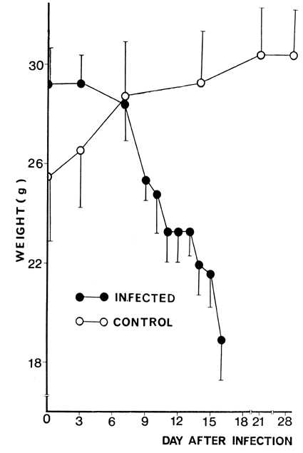
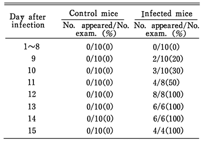
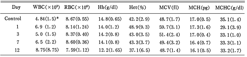
To observe the clinical course and intestinal histopathology in heavy infection of Fibricola seoulensis, an experimental study was performed in mice. Clinical, hematological and histopathological observation was done on 1, 3, 7 and 12 days after experimental infection with l,000 metacercariae. On the 11th day after infection, the mice began to die and all of the infected mice were dead by the 16th day. The infected mice showed gradual weight loss. Occult blood was detected after the 10th day. Diarrhea occurred after the 9th day and was recognized in all of the infected mice on the 12th day. Hemoglobin and mean corpuscular hemoglobin decreased significantly after the 12th day, and mean corpuscular hemoglobin concentration decreased in all infected mice. The histopathological changes were more marked in the duodenum than in the jejunum or ileum. Major changes were villous atrophy and crypt hyperplasia, with decreased villus/crypt ratio, inflammatory cell infiltration and stromal edema. The present results suggest that the cause of death of mice heavily infected with F. seoulensis should be malnutrition and severe fluid loss due to malabsorption, together with intestinal bleeding.
Figures
Fig. 1 Survival rate of mice fed 1,000 metacercariae of F. seoulensis, in comparison with control group.
Fig. 2 Weight change of mice fed 1,000 metacercariae of F. seoulensis, in comparison with control group. (工 ; standard deviation)
Figs. 3-11 Fig. 3. The gross feature of an intestinal segment of a mouse dead on day 16 after infection, showing gross luminal bleeding and worms (arrows).
Fig. 4. Mouse duodenum of control group, showing normal and long, finger-like villi. H-E stain, ×100.
Fig. 5. Duodenal section of a 1-day infected mouse showing a young fluke in the intervillous space, H-E stain, ×100.
Fig. 6. Duodenal section of a 3-day infected mouse showing a worm entrapping the tip of a villus. Destruction of epithelial cells adjacent to the worm, and compression of the villus is conspicious. H-E stain, ×100.
Fig. 7. Duodenal section of a 7-day infected mouse showing villous atrophy, crypt hyperplasia, cellular infiltration and decreased V/C ratio (about 1:1). H-E stain, ×40.
Fig. 8. Duodenal section of a 7-day infected mouse showing a mature worm entrapping the villus, and showing destroyed villi. H-E stain, ×100.
Fig. 9. Jejunal section of a 7-day infected mouse showing stromal edema and decreased V/C ratio. H-E stain, ×40.
Fig. 10. Duodenal section of a 12-day infected mouse showing marked villous atrophy, crypt hyperplasia and reversed V/C ratio (1:2). H-E stain, ×40.
Fig. 11. Ileal section of a 12-day infected mouse showing severe stromal edema and flatting of villi and markedly decreased V/C ratio (2:1 to 1:1). H-E stain, ×100.
Tables
Table 1 Occult blood positive rate in mice fed 1,000 metacercariae of F. seoulensis by infection day
Table 2 Appearance rate of diarrhea in mice fed 1,000 metacercariae of F. seoulensis by infection day
Table 3 Number of recovered worms from each mouse according to duration of survival days
Table 4 Hematological values of mice fed 1,000 metacercariae of F. seoulensis by day
References
1.
Bindseil E, Christensen NO. Thymus-independent crypt hyperplasia and villous atrophy in the small intestine of mice infected with the trematode Echinostoma revolutum. Parasitology 1984;88(Pt 3):431–438.
2.
Chai JY. Seoul J Med 1979;20(2):104–116.
3.
Chai JY, Seo BS, Lee SH. Study On Metagonimus Yokogawai(Katsurada, 1912) In Korea Vii. Susceptibility Of Various Strains Of Mice To Metagonimus Infection And Effect Of Prednisolone. Korean J Parasitol 1984;22(2):153–160.
4.
Cho SY, et al. Chung-Ang J Med 1983;8(1):13–28.
5.
Erasmus DA, Ohman C. Electron microscope studies of the gland cells and host-parasite interface of the adhesive organ of Cyathocotyle bushiensis Khan, 1962. J Parasitol 1965;51(5):761–769.
6.
Ferguson A, Jarrett EE. Hypersensitivity reactions in small intestine. I Thymus dependence of experimental 'partial villous atrophy'. Gut 1975;16(2):114–117.
8.
Hong ST. Studies On Intestinal Trematodes In Korea: VII. Growth, Development And Recovery Of Fibricola Seoulensis From Experimentally Infected Rats And Mice. Korean J Parasitol 1982;20(2):112–121.
9.
Hong ST, Chai JY, Lee SH. Ten human cases of Fibricola seoulensis infection and mixed one with Stellantchasmus and Metagonimus. Korean J Parasitol 1986;24(1):95–97.
10.
Hong ST, Cho TK, Hong SJ, Chai JY, Lee SH, Seo BS. Fifteen human cases of Fibricola seoulensis infection in Korea. Korean J Parasitol 1984;22(1):61–65.
11.
Huffman JE, Michos C, Fried B. Clinical and pathological effects of Echinostoma revolutum (Digenea: Echinostomatidae) in the golden hamster, Mesocricetus auratus. Parasitology 1986;93(Pt 3):505–515.
12.
Lee SH, Yoo BH, Hong ST, Chai JY, Seo BS, Chi JG. A histopathological study on the intestine of mice and rats experimentally infected by Fibricola Seoulensis. Korean J Parasitol 1985;23(1):58–72.
13.
Liebman WM, Thaler MM, DeLorimier A, Brandborg LL, Goodman J. Intractable diarrhea of infancy due to intestinal coccidiosis. Gastroenterology 1980;78(3):579–584.
14.
Manson-Smith DF, Bruce RG, Parrott DM. Villous atrophy and expulsion of intestinal Trichinella spiralis are mediated by T cells. Cell Immunol 1979;47(2):285–292.
15.
Milner PF, Irvine RA, Barton CJ, Bras G, Richards R. Intestinal malabsorption in Strongyloides stercoralis infestation. Gut 1965;6(6):574–581.
16.
Rho IH, et al. Chung-Ang J Med 1984;9(1):67–78.
17.
Ruitenberg EJ, Leenstra F, Elgersma A. Thymus dependence and independence of intestinal pathology in a Trichinella spiralis infection: a study in congenitally athymic (nude) mice. Br J Exp Pathol 1977;58(3):311–314.
18.
Seo BS, et al. Seoul J Med 1986;27(2):125–134.
19.
Seo BS, Lee SH, Hong ST, Hong SJ, Kim CY, Lee HY. Studies On Intestinal Trematodes In Korea: V. A Human Case Infected By Fibricola Seoulensis (Trematoda: Diplostomatidae). Korean J Parasitol 1982;20(2):93–99.