Warning: mkdir(): Permission denied in /home/virtual/lib/view_data.php on line 81
Warning: fopen(upload/ip_log/ip_log_2024-04.txt): failed to open stream: No such file or directory in /home/virtual/lib/view_data.php on line 83
Warning: fwrite() expects parameter 1 to be resource, boolean given in /home/virtual/lib/view_data.php on line 84 Histopathological and serological observations on experimental anisakiasis of rabbits
Histopathological and serological observations on experimental anisakiasis of rabbits
Sung Tae Hong and Soon Hyung Lee
Department of Parasitology and Institute of Endemic Diseases, College of Medicine, Seoul National University, Seoul 110, Korea.
Abstract
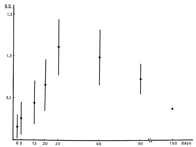
This study was performed to observe histopathological changes and serological reactions in chronic anisakiasis of rabbits. Each rabbit was infected per os with 30 larvae of Anisakis type I. Their sera were collected chronologically and the rabbits were killed for histopathological examination, 3, 13, 20, 30, 60, 90 and 150 days after the infection. The results were summarized as below. Most of the larvae were recovered from the stomach, but a few from the omentum, intestine, mesentery and abdominal wall. The recovery rates and distribution of worms by organ were not differed by duration of infection. Histologically the lesion was abscess type on 13 days, i.e., the dead worms were surrounded by fibrinous exudate, histiocytes and thick zone of numerous inflammatory cells. After 30 days, histiocytes were found to invade the worms and the lesion was changing into abscess-granulomatous type. Also a calcified worm was found on the 30th day. After then the worms were observed to be dissolved slowly until 90 days. On 150 day, only one calcified worm was observed. The levels of serum IgG antibody by ELISA reached their maximum 30 days after the infection. After then, it decreased slowly until 150 days after the infection. Above serological and histopathological findings indicated that antigenic stimulation from degenerating Anisakis larvae was the greatest during the first 30 days after infection. This period was corresponding with the beginning of worm resolution or calcification. Serologic test by ELISA would be a valuable tool for confirming chronic anisakiasis.
Figures
Fig. 1 IgG antibody levels (O.D. at 492nm) by ELISA in sera of experimental rabbits by duration of infection.
Fig. 2 Mean (±standard deviation) of the optical densities by ELISA during the course of experimental anisakiasis.
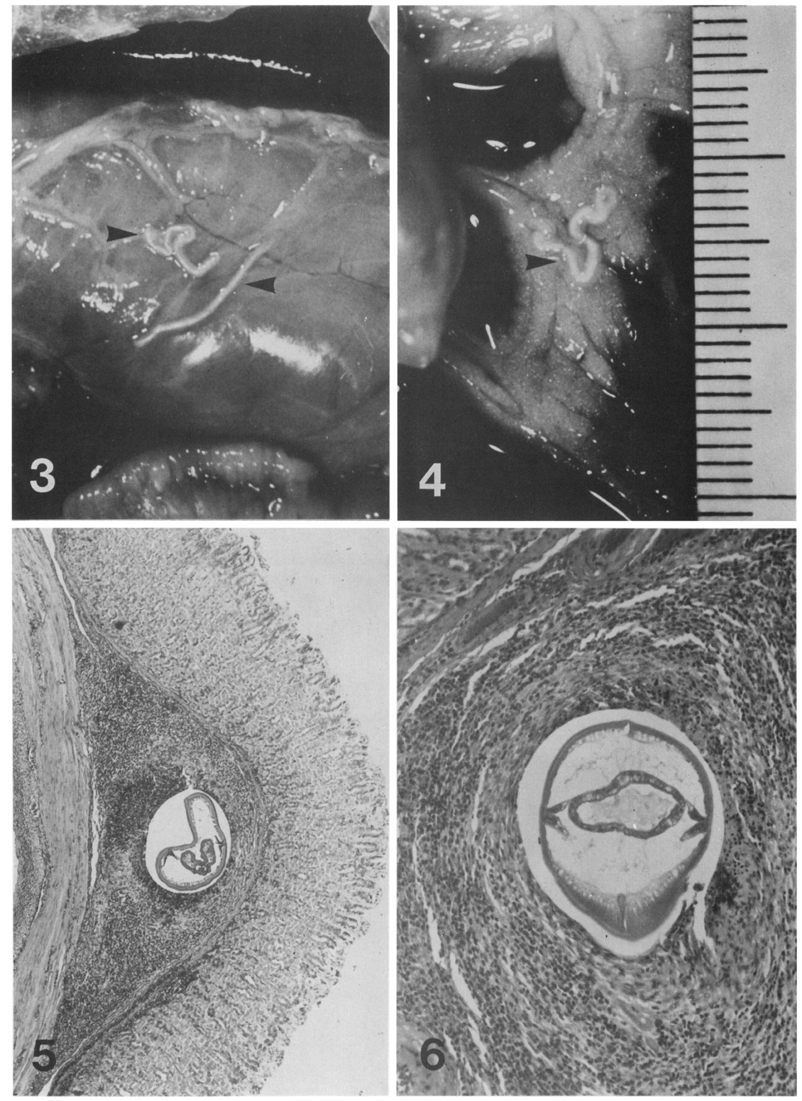
Figs. 3-6 Fig. 3. Two Anisakis larvae (arrow heads) on the surface of the stomach, 30 days after infection.
Fig. 4. A calcified, chalky white and stony hard larva (arrow head) was found in omentum, 60 days after infection.
Fig. 5. A section of Anisakis larva within abscess at submucosa of stomach, 13 days after infection, HE stained, ×40.
Fig. 6. A sectioned larva which preserved its structures was surrounded by histiocytes, fibroblasts and inflammatory cells in submucosa of stomach, 13 days after infection, HE stained, ×100.
Figs. 7-10 Fig. 7. Sections of a necrotizing worm in fibrinous exudate, which is surrounded by histiocytes and by numerous inflammatory cells in stomach, 13 days after infection, HE stained, ×40.
Fig. 8. High power view of a larva surrounded by fibrinous exudate, histiocytes and inflammatory cells in stomach, 13 days after infection, HE stained, ×100.
Fig. 9. Sections of a larva in omentum surrounded by exudate, and inflammatory cells made a mass, 13 days after infection, HE stained, ×40.
Fig. 10. High power view of Fig. 9. showing neighboring fibrinous exudate and the cells (neutrophils, eosinophils, histiocytes and fibroblasts), HE stained, ×100.
Figs. 11-14 Fig. 11. A necrotizing worm in peritoneum, 20 days after infection, with layers of histiocytes, inflammatory cells and fibrosis, HE stained, ×100.
Fig. 12. A shrunk larva surrounded by fibrosis in mesentery, 20 days after infection. The bulk of inflammatory cells diminished, HE stained, ×100.
Fig. 13. A larva was attached by host cells in stomach wall, 30 days after infection, HE stained, ×100.
Fig. 14. High power view of Fig. 13, host cells attaching the surface of worm. Foamy histiocytes and pyknotic debris were seen nearby, HE stained, ×400.
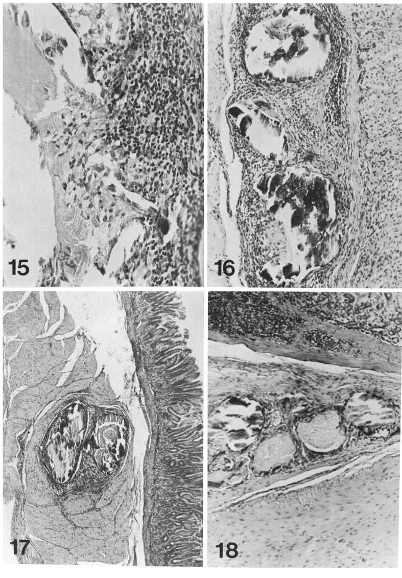
Figs. 15-18 Fig. 15. Worm debris scavenged by histiocytes in stomach wall, 60 days after infection, HE stained, ×200.
Fig. 16. Sections of a calcified worm surrounded by fibrosis. Inflammatory cells were much less, 90 days after infection, HE stained, ×100.
Fig. 17. A calcified worm in the muscle layer of stomach enclosed by thin fibrous tissue, 90 days after infection, HE stained, ×40.
Fig. 18. A calcified worm in the stomach wall with surrounding fibrosis, 150 days after infection, HE stained, ×40.
Tables
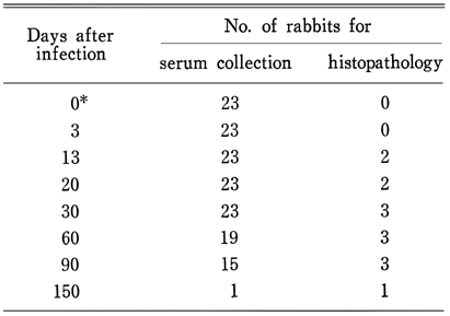
Table 1 The number of rabbits used by the duration of infection
Table 2 Numbers of recovered worms in experimental anisakiasis of rabbits by location
References
1.
Cho SY, et al. Seoul J Med 1980;21(2):203–208.
2.
Choi WJ, et al. Seoul J Med 1984;25(4):569–577.
3.
Choi WY, Yoo JE, Nam HW, Choi HR. [Purification of antigenic proteins of Paragonimus westermani and their applicability to experimental cat paragonimiasis]. Korean J Parasitol 1986;24(2):177–186.
4.
Desowitz RS, Raybourne RB, Ishikura H, Kliks MM. The radioallergosorbent test (RAST) for the serological diagnosis of human anisakiasis. Trans R Soc Trop Med Hyg 1985;79(2):256–259.
5.
Jeong JS, et al. Inje Med J 1984;5(3):359–367.
6.
Kim CH, Chung BS, Moon YI, Chun SH. [A case report on human infection with Anisakis sp. in Korea]. Korean J Parasitol 1971;9(1):39–43.
7.
Lee A, et al. Korean J Pathol 1985;19(4):463–467.
8.
Lee CY, et al. Korean J Med Technologists 1986;18(1):171–176.
9.
Lee KH, et al. Korean J Int Med 1981;24(12):1220–1227.
10.
McLaren M, Draper CC, Roberts JM, Minter-Goedbloed E, Ligthart GS, Teesdale CH, Amin MA, Omer AH, Bartlett A, Voller A. Studies on the enzyme linked immunosorbent assay (ELISA) test for Schistosoma mansoni infections. Ann Trop Med Parasitol 1978;72(3):243–253.
11.
Oshima T. Prog of Med Parasit in Japan 1972;4:305–393.
12.
Oshima T. Anisakiasis - is the sushi bar guilty. Parasitol Today 1987;3(2):44–48.
13.
Oyanagi T. Jpn J Parasitol 1967;16(6):470–493.
14.
Paik AL, et al. Korean J Path 1984;18(4):453–456.
15.
Seo BS, Chai JY, Lee SH, Hong ST, Seo JW, Noh SH. A human case infected by the larva of Terranova type A in Korea. Korean J Parasitol 1984;22(2):248–252.
16.
Smith JW, Wootten R. Anisakis and anisakiasis. Adv Parasitol 1978;16:93–163.
17.
Suzuki T, Sato Y, Yamashita T, Sekikawa H, Otsuru M. Anisakiasis: preparation of a stable antigen for indirect fluorescent antibody test. Exp Parasitol 1974;35(3):418–424.
18.
Takahashi S, et al. Jpn J Parasit 1986;35(2):86–87.
19.
van Thiel P, Kuipers FC, Roskam RT. A nematode parasitic to herring, causing acute abdominal syndromes in man. Trop Geogr Med 1960;12:97–113.
20.
Yoshimura H, Akao N, Kondo K, Ohnishi Y, Funaoka Y, Yamane K. [Two cases of extra-gastrointestinal anisakiasis and evaluation of immunodiagnosis (author\'s transl)]. Rinsho Byori 1980;28(7):708–712.