Warning: mkdir(): Permission denied in /home/virtual/lib/view_data.php on line 81
Warning: fopen(upload/ip_log/ip_log_2024-04.txt): failed to open stream: No such file or directory in /home/virtual/lib/view_data.php on line 83
Warning: fwrite() expects parameter 1 to be resource, boolean given in /home/virtual/lib/view_data.php on line 84 Ultrastructural studies on the surface of Paragonimus westermani metacercaria
Ultrastructural studies on the surface of Paragonimus westermani metacercaria
Kyong Min Kim,Myoung Hee Ahn and Duk Young Min
Department of Parasitology, Hanyang University Medical College, Seoul 133, Korea.
Abstract
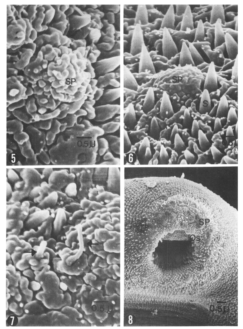
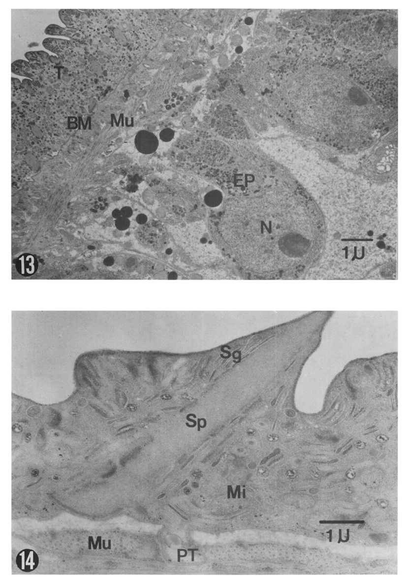
The present study was undertaken to demonstrate the surface structure of Paragonimus westermani metacercaria in Korea with special reference to the distribution of sensory papillae. Metacercariae were isolated from crayfish, one of the second intermediate host of P. westermani in Bogil island, Chollanam-do (Province), Korea, where has been known as an endemic area of human paragonimiasis. Isolated metacercariae were excysted and examined with light, scanning and transmission electron microscopes for morphological features. On the surface of metacercariae, three types of sensory papillae were identified. Large domed papillae (3-5 µm), which were covered with wrinkled plasma membrane of the worm, were distributed on the oral and ventral suckers only. On the oral sucker, these large domed papillae were 12-13 in number. On the other hand large domed papillae on the ventral sucker were constantly 6 in number and hexagonal in distribution. Small domed papillae (2-3 µm), of which surface was more smooth than those of large ones, were distributed symmetrically on the ventral (30-32 pairs) and dorsal surfaces (40-42 pairs). Ciliated papillae (0.8-1.5 µm) were observed about 5-6 in number around the oral sucker and 3-5 pairs each on the ventral and dorsal surface of the body. Single pointed spines covered the entire surface of the body except around the excretory pore. Spines on the anterior part of the body were 0.9-2.0 µm in length and 45-55/100 square µm in number, and were gradually reduced in length (0.4-1.4 µm) and in number (12-27/100 square µm) toward the posterior part. The body wall of P. westermani metacercariae was consisted with anucleated syncytium layer, fibrous interstitial layer and musclar layer. In the anucleated syncytium, biconcave (0.15-0.55 µm) and spherical (0.08-0.16 µm) secretory granules, which were transferred from epidermal cells via protoplasmic tubules, mitochondria and ribosomes, were observed. Spines originated around the basement membrane protruded externally. Epidermal cells were consisted with a nucleus and a cytoplasm, and connected to syncytium with protoplasmic tubules. In the cytoplasm many secretory granules, mitochondria, Golgi complex, endoplasmic reticula, ribosomes and lipid droplets were observed.
Figures
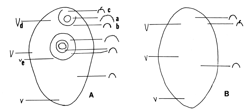
Diagram 1 Schematic diagram of tegumental differentiation of excysted metacercaria of P. westermani, ventral (A) and dorsal (B) view.
(a: large domed papillae, b: small domed papillae, c: ciliated papillae, d: large spine, e: small spine)
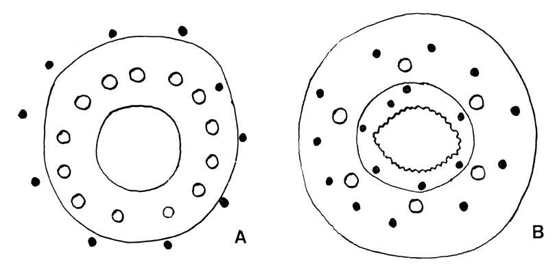
Diagram 2 Schematic diagram of sensory papillae around oral(A) and ventral suckers (B).
(◦ : large domed papillae, • : small domed papillae)
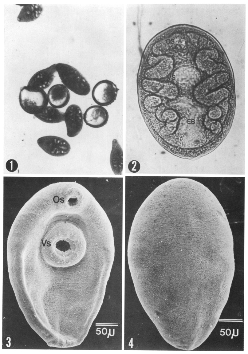
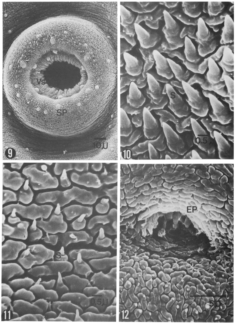
Figs. 1-4 Fig. 1. Excysted metacercariae of Paragonimus westermani.
Figs. 15-16 Fig. 15. Large domed sensory papilla on the ventral sucker of P. westermani metacercaria (DB: dense body of papilla, Sp: spine) (×10,000).
Fig. 16. Small domed sensory papilla on the dorsal surface of P. westermani metacercaria (Sp: spine, V: vesicle of papilla) (×12,500).
Tables
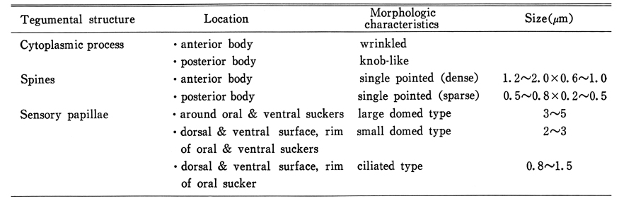
Table 1 Surface findings of excysted P. westermani metacercaria by SEM
Table 2 Number of sensory papillae of excysted P. westermani metacercaria
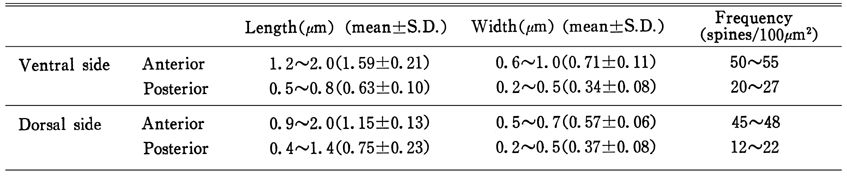
Table 3 Size and frequency of spines of excysted P. westermani metacercaria
References
1.
Aji T, et al. . Jpn J Parasit 1984;33:15–21.
2.
Bennett CE. Surface features, sensory structures, and movement of the newly excysted juvenile Fasciola hepatica L. J Parasitol 1975;61(5):886–891.
3.
Bennett CE. Scanning electron microscopy of Fasciola hepatica L. during growth and maturation in the mouse. J Parasitol 1975;61(5):892–898.
4.
Burton PR. The Ultrastructure Of The Integument Of The Frog Lung-Fluke, Haematoloechus Medioplexus (Trematoda: Plagiorchiidae). J Morphol 1964;115:305–317.
5.
Burton PR. The ultrastructure of the integument of the frog bladder fluke, Gorgoderina sp. J Parasitol 1966;52(5):926–934.
6.
Choi WY, Yoo JE. [Ultrastructure of the integument of adult Paragonimus westermani]. Korean J Parasitol 1985;23(1):111–122.
7.
Chung PR. Yonsei Rep Trop Med 1983;14:44–65.
8.
Fujino T, Ishii Y, Cho DW. Surface ultrastructure of the tegument of Clonorchis sinensis newly excysted juveniles and adult worms. J Parasitol 1979;65(4):579–590.
9.
Habe S, et al. Jpn J Parasit 1982;31:275–280.
10.
Habe S, et al. Jpn J Parasit 1982;31:27–32.
11.
Higo H, et al. Jpn J Parasit 1984;33:421–427.
12.
Lee DL. The structure and composition of the helminth cuticle. Adv Parasitol 1966;4:187–254.
13.
Lee SH, Hong ST, Seo BS. [A Study On The Fine Tegumental Structures Of The Metacercaria And Juvenile Stages Of Clonorchis Sinensis]. Korean J Parasitol 1982;20(2):123–132.
14.
Lee SH, Seo BS, Chai JY, Hong SJ. [Study on Metagonimus yokogawai(Katsurada, 1912) in Korea VII. Electron microscopic observation on the tegumental structure]. Korean J Parasitol 1984;22(1):1–10.
15.
Lumsden RD. Surface ultrastructure and cytochemistry of parasitic helminths. Exp Parasitol 1975;37(2):267–339.
16.
Miyazaki I. Med Bull Fukuoka Univ 1978;5:251–263.
17.
Seo BS, Lee SH, Chai JY, Hong ST, Hong SJ. [Studies on intestinal trematodes in Korea X. Scanning electron microscopic observation on the tegument of Fibricola seoulensis]. Korean J Parasitol 1984;22(1):21–29.