Warning: mkdir(): Permission denied in /home/virtual/lib/view_data.php on line 81
Warning: fopen(upload/ip_log/ip_log_2024-04.txt): failed to open stream: No such file or directory in /home/virtual/lib/view_data.php on line 83
Warning: fwrite() expects parameter 1 to be resource, boolean given in /home/virtual/lib/view_data.php on line 84 Five human cases of Diphyllobothrium latum infection through eating raw flesh of redlip mullet, Liza haematocheila: Article in Korean
Five human cases of Diphyllobothrium latum infection through eating raw flesh of redlip mullet, Liza haematocheila: Article in Korean
P R Chung,*1W M Sohn,2Y Jung,1S H Pai,3 and M S Nam4
1Department of Parasitology, Inha University College of Medicine, Inchon 402-751, Korea.
Received September 18, 1997; Accepted October 22, 1997.
Abstract
A total of 32 human diphyllobothriasis cases have been reported so far in Korea, excluding 21 egg-positive cases from stool examinations. Authors experienced five more human cases of Diphyllobothrium latum infection, especially infected due to eating raw flesh of redlip mullet, Liza haematocheila. Five cases were neighbors residing in the Puchon area, Kyonggi-do, who ate raw mullets (L. haematocheila) in a party in February 1996. The mullets were purchased at the Noryangjin fisheries market in Seoul. All of cases (2 males and 3 females) were 35 to 43 years old and healthy with the body weight range of 56-62 kg. They complained about gastrointestinal trouble and abdominal discomfort, but were in normal ranges of their hematology and urinalysis data. None revealed any sign of anemia. The patients experienced natural discharge of a chain of segments before, and showed diphyllobothriid eggs in their stool specimens when they visited our laboratory. They were administered with praziquantel (15 mg/kg of body weight) and 30 g of magnesium sulfate as a purgative. Two whole worms with the scolices (310-340 cm in length; 8-13 mm in width) were expelled each from two out of five cases after anthelmintic treatment, and the others expelled the parts of strobilae without scolex. The worms were identified as D. latum, based on the following biological characters: external morphologies, coiling of uterus, the number of uterine loops, position of genital opening, morphologies of cirrus, cirrus sac and seminal vesicle on the histological sections, position of vagina and uterine pore, and microscopical and SEM morphologies of the eggs.
Figures
Figs. 1-9 Fig. 1. A whole strobila of D. latum with scolex, collected from case 1(320 cm in length). Fig. 2. Close-up view of gravid segments of D. latum showing dark uterine fields. Fig. 3. Two gravid segments of D. latum showing rosette-shaped uteri, stained with acetocarmine (Go, genital opening; U, uterus). Fig. 4. A scolex of D. latum from case 3 (B, bothrium). Fig. 5. An egg of D. latum from case 5 (Ak, abopercular knob; Op, operculum). Fig. 6. SEM view of a D. latum egg showing inconspicuous tiny pits on the egg shell (×5,000) (Op, operculum; P, pit). Fig. 7. Longitudinal section of a mature proglottid, showing the complete cirrus sac, cirrus, seminal vesicle and uterus (c, cirrus; Cs, cirrus sac; Sv, seminal vesicle; U, uterus; Vf, vitelline follicles). Fig. 8. SEM view of the genital atrium area of a mature proglottid collected from case 5(×50) (C, cirrus; Up, uterine pore). Fig. 9. Magnification of the genital atrium, showing the genital papillae on the genital atrium (×100) (C, cirrus; Gp, genital papillae; Up, uterine pore).
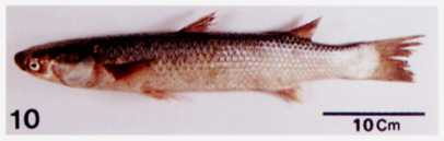
Fig. 10 A redlip mullet, Liza haematocheila (Temminck et Schlegel), probable infection source of present cases.
References
1.
Ahn YK, et al. Korean J Biomed Lab Sci 1996;2(2):283–288.
2.
Brooke MM, et al. US Armed Forces Med J 1956;7:708–714.
3.
Cho SY, et al. Seoul J Med 1971;12(3):157–160.
4.
Chyu I, et al. Theses of Catholic Med Coll 1965;9:159–172.
5.
Eguchi S. Prog Med Parasit in Japan 1973;5:129–144.
6.
Hara C, et al. Chosen Igakkai Zasshi 1923;48:112–122.
7.
Hotta J, et al. Jpn J Parasitol 1978;27(4):357–368.
8.
Kobayashi H. Jpn Med World 1925;5(1):9–16.
9.
Kojima R, et al. Chosen Igakkai Zasshi 1919;26:42–86.
10.
Lee SH, et al. Seoul J Med 1988;29:391–395.
11.
Lee SH, Chai JY, Seo M, Kook J, Huh S, Ryang YS, Ahn YK. Two rare cases of Diphyllobothrium latum parvum type infection in Korea. Korean J Parasitol 1994;32(2):117–120.
12.
Lee SH, Seo BS, Chai JY, Hong ST, Hong SJ, Cho SY. [Five Cases Of Diphyllobothrium Latum Infection]. Korean J Parasitol 1983;21(2):150–156.
13.
Min DY. Cestode infections in Korea. Korean J Parasitol 1990;28 Suppl:123–144.
14.
Min DY, Ahn MH, Kim KM, Kim CW. [Intestinal parasite survey in Seoul by stool examination at Hanyang University Hospital]. Korean J Parasitol 1986;24(2):209–212.
15.
Petruschewsky GK, et al. Tropenhyg 1933;37(6):307–315.
20.
Yamane Y, et al. Shimane J Med Sci 1986;10:29–48.
21.
Yamane Y, et al. Jpn J Parasitol 1981;30(2):101–111.
22.
Yamane Y, et al. Jpn J Parasitol 1983;32(1):13–25.
24.
Yokogawa M, et al. Jpn J Parasitol 1979;28(3):133–138.