A Case of Echinostoma cinetorchis (Trematoda: Echinostomatidae) Infection Diagnosed by Colonoscopy
Article information
Abstract
Human cases of echinostomiasis have been sporadically diagnosed by extracting worms in the endoscopy in Korea and Japan. Most of these were caused by Echinostoma hortense infection. However, in the present study, we detected 2 live worms of Echinostoma cinetorchis in the ascending colon of a Korean man (68-year old) admitted to the Gyeongsang National University Hospital with complaint of intermittent right lower quadrant abdominal pain for 5 days. Under colonoscopy, 1 worm was found attached on the edematous and hyperemic mucosal surface of the proximal ascending colon and the other was detected on the mid-ascending colon. Both worms were removed from the mucosal surface with a grasping forceps, and morphologically identified as E. cinetorchis by the characteristic head crown with total 37 collar spines including 5 end-group ones on both sides, disappearance of testes, and eggs of 108×60 µm with abopercular wrinkles. The infection source of this case seems to be the raw frogs eaten 2 months ago. This is the first case of endoscopy-diagnosed E. cinetorchis infection in Korea.
INTRODUCTION
Echinostomes (Family Echinostomatidae) are parasitic intestinal flukes which infect a wide range of vertebrates including humans [1]. Among the species, Echinostoma. cinetorchis was first discovered from rats in Japan, and then from dogs and rats in Taiwan and the Republic of Korea (=Korea) [2,3,4,5,6]. This fluke was also found in China [7] and Vietnam [8,9]. Human infections with this fluke were first reported in Japan and then in Korea [10,11,12,13,14,15,16]. This human intestinal fluke is morphologically characterized by a head crown with 37-38 collar spines including 5 end group ones on each side, and abnormal location and/or disappearance of testis [1,2].
Echinostomiasis is a foodborne intestinal trematodiasis caused by a total of 20 species belonging to 9 genera in the Family Echinostomatidae [1]. It is endemic in southeast Asia and the Far East, i.e., Malaysia, Philippines, Indonesia, Thailand, Lao PDR, Cambodia, mainland China, Taiwan, India, and Korea [17,18,19,20]. In the genus Echinostoma, more than 7 species are known to be zoonotic, i.e., E. angustitestis, E. cinetorchis, E. ehinatum, E. hortense, E. ilocanum, E. macrorchis, and E. revolutum [1]. Among them, Echinostoma hortense is the predominant species with several endemic foci in Korea [15,16,21,22,23]. Clinical cases of E. hortense infection have been sporadically diagnosed by the recovery of adult worms in gastroduodenal endoscopy [24,25,26,27,28]. However, human cases of E. cinetorchis infection were not many and never diagnosed through endoscopy. In this study, we report a clinical case of E. cinetorchis infection diagnosed by colonoscopy.
CASE RECORD
A 68-year-old Korean man, residing in Geoje-si, Gyeongsangnam-do, was admitted to the Gyeongsang National University Hospital with complaint of intermittent right lower quadrant abdominal pain for 5 days. He underwent surgical resection of esophageal tumor; thereafter, he took chemotherapy with R-CHOP regimen of 6 cycles as a diagnosis of diffuse large B-cell lymphoma 4 years ago. Eosinophilia was found at the time of esophageal surgery. He also underwent laparoscopic cholecystectomy due to acute cholecystitis 1 year ago. Several times fecal examinations were performed, but the results were negative for occult blood and parasites. He had taken praziquantel 2 times; however, eosinophilia persisted until recent days. He often ate raw beef and sliced raw fish. In particular, he had eaten raw frogs 2 months ago.
At the time of admission, his body temperature was 36.2℃ and blood pressure was 110/70 mmHg. There were no abnormal findings on physical examination. Laboratory data included the followings: hemoglobin 14.7 g/dl (normal: 13.0-17.0), hematocrit 30.3% (39-52%), platelet 220,000/mm3 (130,000-400,000/mm3), white blood cell count 4,870/mm3 (4,000-10,000/mm3), and eosinophil 20.7% (1-5%). Liver function tests, serum electrolytes, and urinalysis were within normal limit. Fecal examination was negative for occult blood and parasites. Abdominal CT showed mild intrahepatic and common bile duct dilatation and diverticulum-like lesion at the ascending colon with adjacent mesenteric infiltration. Colonoscopy revealed 2 live worms with regional mucosal erythematous swellings in the ascending colon. One worm was found in the proximal ascending colon and the other was in the mid-ascending colon. The mucosal surface adjacent to the worms was edematous and hyperemic (Fig. 1). However, no diverticulum was found. The worms wriggled and moved in the manner of elongation and shortening on stimulation. Both worms were removed from the mucosal surface with a grasping forceps. The live parasites were transferred to the Department of Parasitology and Tropical Medicine, Gyeongsang National University for the identification of worms. Microscopic examinations of endoscopic biopsy specimens revealed chronic non-specific colitis with lymphoid aggregation in the colonic mucosa.
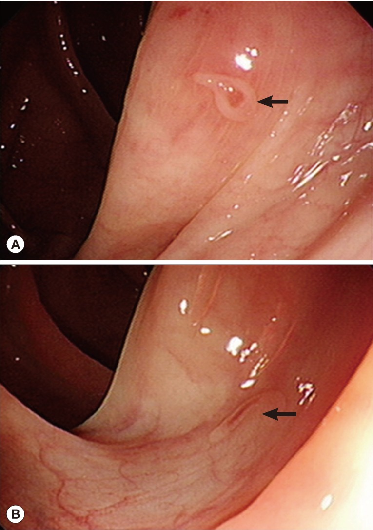
Colonoscopic views of the patient. (A) A bending worm (arrow) is attached to the hyperemic, edematous mucosal surface of the proximal ascending colon. (B) Another elongated worm (arrow) is seen on the mid-ascending colon. They were wriggling and moving in the manner of elongation and shortening by the stimuli of the grasping forceps.
The patient was treated with praziquantel, 25 mg/kg, given orally 3 times in a day, and thereafter his symptoms disappeared. The number of white blood cells was 4,990/mm3 and the percentage of eosinophil decreased markedly to 5.4% in 2 months.
The recovered worms were morphologically observed under a light microscope with a micrometer after fixation with 10% formalin under the cover slip pressure and staining with Semichon's acetocarmine (Fig. 2). They were elongated, spindle shaped, and 5,600 µm (in average) long and 1,563 µm wide (maximum) at the ovarian level. Its characteristic head crown was reniform (180 by 438 µm), and had 37 collar spines in 2 rows including 5 end-group spines on each side. The oral sucker was transversely elliptical (170 by 228 µm). The ventral sucker was well developed (615 by 580 µm) and located at 1,300 µm from the anterior end. The pharynx was muscular (195 by 143 µm). The esophagus was slender (425 µm long). Two ceca was bifurcated in front of the cirrus sac and extended to near the posterior end of the body. The cirrus sac was oval (310 µm long and 175 µm wide), located median between the preacetabular and cecal bifurcation. The ovary was transversely oval (235 by 380 µm) and located equatorial in the median line. The Mehlis' gland was conspicuous and located immediately postovarian. They all had no testes. Uterine coils with numerous eggs were distributed between the ventral sucker and ovary. Vitellaria were follicular and distributed mainly at lateral fields from behind the posterior margin of the ventral sucker to the posterior end. Intrauterine eggs were operculated and yellowish (av. 108 by 60 µm).
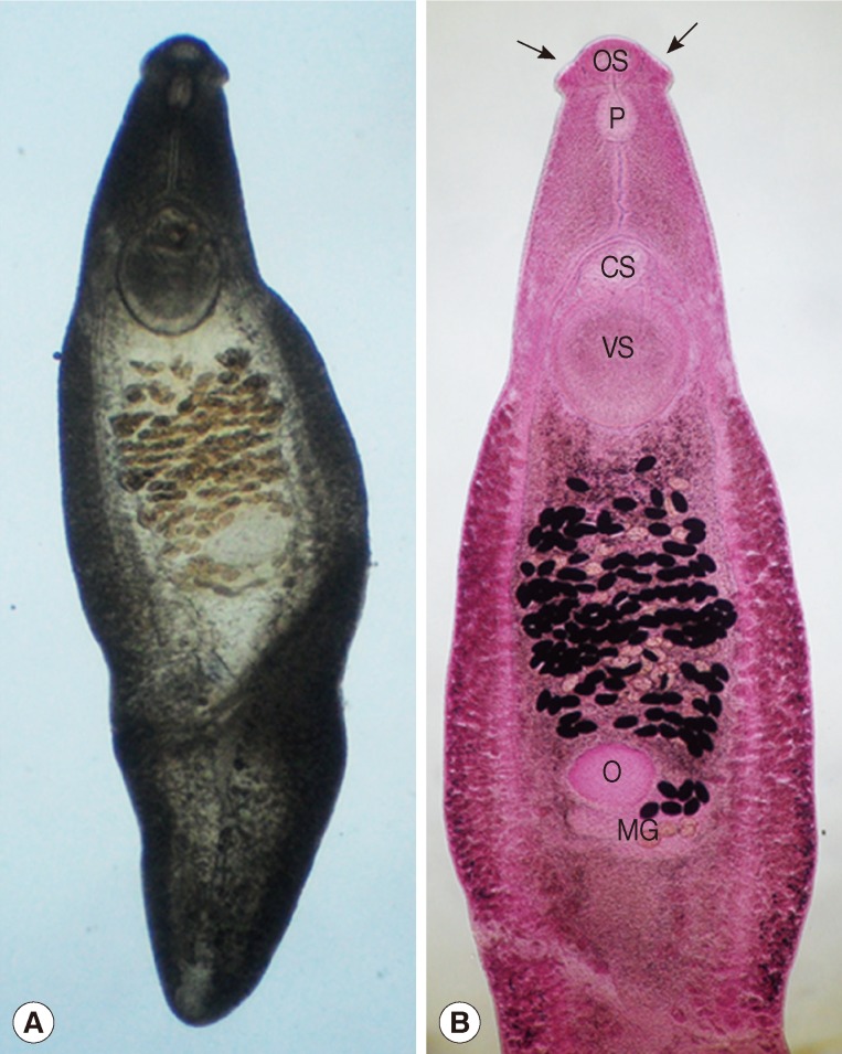
Two adult Echinostoma cinetorchis recovered in the colon of the present case. (A) An unstained worm fixed in 10% formalin showing the complete feature after removing with a grasping forceps. (B) Another worm stained with Semichon's acetocarmine, which was broken in the posterior middle portion during the extracting process, but reveals the typical morphologies, i.e., a reniform head crown (arrows) around the oral sucker (OS), muscular pharynx (P), well-developed ventral sucker (VS), oval-shaped cirrus sac (CS), transversely elliptical ovary (O), and conspicuous Mehlis' gland (MG), but no testes in the postovarian region. The worms were characterized by the head crown equipped with 37 collar spines including 5 end-group spines on each side, and no testis in the postovarian and the posterior middle portion of worms. Scale bar is 1,000 µm.
DISCUSSION
The present case is the first E. cinetorchis infection diagnosed by colonoscopy in the literature. Until now, in Korea, a total of 6 human E. cinetorchis infections have been reported based on adult worm recovery [12,13,14,15,16]. With regard to E. hortense, the dominant species, it is known to be endemic in some areas of Eumseong-gun (Chungcheongbuk-do), Yeongwol-gun (Gangwon-do), Cheongsong-gun (Gyeongsangbuk-do) and Geochang-gun (Gyeongsangnam-do) in Korea [15,16,21,22,23]. Several clinical cases of E. hortense were sporadically diagnosed by extracting worms through gastroduodenal endoscopy [24,25,26,27,28].
In our case, the worm recovery from the colon is quite interesting, considering that the main habitat of E. cinetorchis is the small intestine as shown in experimental and natural definitive hosts [5,6,29,30]. It is unknown whether the colon is a favored habitat of E. cinetorchis worms in the human host. Recovery of worms in other endoscopy and autopsy cases will be helpful to determine it, and clinicians should pay attention to this fluke in colonoscopic examinations.
Freshwater snails, Hippeutis cantori and Segmentina hemisphaerula, were experimentally confirmed as the first and second intermediate hosts of E. cinetorchis. Other freshwater snails, including Radix auricularia coreana, Physa acuta, Austropeplea ollula, Corbicula fluminea, Pisidium coreanum, and Cipangopaludina chinensis malleata, were reported to be the second intermediate hosts [1,29,30]. Tadpoles and frogs of Rana spp. and the loach, Misgurnus anguillicaudatus, were also proved to harbor the metacercariae [1,31]. Accordingly, the infection source of this case may be raw frogs under the circumstances of the past history of the patient and the biological characteristics of this fluke.
Generally, a single dose of 10 mg/kg praziquantel is recommended as the regimen for the treatment of intestinal fluke infections. In the present study, the patient was treated with praziquantel, 25 mg/kg, given orally 3 times in a day. This regimen is equal to the treatment regimen for clonorchiasis and may be too much for intestinal trematodes. However, this high dose of praziquantel may kill intestinal trematodes better.
Notes
We have no conflict of interest related to this study