Molecular Characterization of Enterocytozoon bieneusi in Domestic Rabbits (Oryctolagus cuniculus) in Northeastern China
Article information
Abstract
A study of 426 rabbits from 3 cities in Jilin province (Changchun City and Jilin City) and Liaoning province (Shenyang City) was conducted between May and June 2015. The overall prevalence of E. bieneusi in rabbits was 0.94% (4/426), with 0% (0/116), 1.72% (3/174), and 0.74% (1/136) in Jilin, Changchun, and Shenyang City, respectively. Only 3 farms (farm 1 and farm 3 in Changchun City, farm 8 in Shenyang City) were PCR-positive for E. bieneusi. Moreover, rabbits of more than 6 months (1.72%) had the highest E. bieneusi prevalence, followed by rabbits of 4-6 months (1.26%), 2-3 months (0.58%), and less than 1 month (0%). Analysis of ITS gene of E. bieneusi suggested that all 4 E. bieneusi isolates were genotype D, and were classified as group 1a. The present results first demonstrated the existence of zoonotic E. bieneusi in domestic rabbits in China. Effective control measures should be implemented to prevent E. bieneusi infection in domestic rabbits, other animals, and humans.
China has a long history of breeding rabbits which have a close relationship with humans. With the improvement of living standards, rabbit meat became widely used for consumption. Thus, more people have pay attention to rabbit industry. Therefore, evaluating how pathogens influence animal health is essential to rabbit industry.
It is well known that the rabbit is a major host for many pathogens including microsporidia [1,2]. However, information regarding the prevalence and genotypes of Encephalitozoon bieneusi in China is scarce. The objectives of the present study were to estimate the prevalence of E. bieneusi in rabbits and to identify their genotypes in China. Moreover, the present study also aimed to provide the foundation data for further study of E. bieneusi genotypes distribution in China.
A total of 426 fecal samples were collected from Jilin (n=290) and Liaoning (n=136) province between May and June 2015. At the time of sampling, all the rabbits were in good health. Fresh fecal sample was collected from each animal using sterile disposal PE gloves after defecation onto the ground. All samples were sent to the laboratory, and then genomic DNA was extracted using an EZNAR Stool DNA kit (OMEGA Biotec, Madison, Wisconsin, USA) according to the manufacturer instructions, immediately. All the extracted DNA samples were stored at -20˚C until PCR analysis. Information concerning gender, geographic origin, and age were obtained by a questionnaire.
E. bieneusi genotypes were determined by nested PCR of the internal transcribed spacer (ITS) region of the ribosomal RNA (rRNA) gene [3]. PCR reaction (25 μl) was composed of 200 μM each deoxy-ribonucleoside triphosphate (dNTP), 1×Ex Taq buffer (Mg2+ free), 0.4 μM of each primer, 0.625 U of Ex Taq DNA polymerase (TAKARA, Kyoto, Japan), 2 mM MgCl2, and 2 μl of DNA template. The cycling conditions were denatured at 95˚C for 5 min, and then 35 cycles of 45 sec at 94˚C, 45 sec at 55˚C, and 1 min at 72˚C, followed by 72˚C for 10 min. Both positive and negative controls were included in each test. All the PCR products were electrophoresed on a 2% agarose gel, stained with GoldView™ (Solarbio, Beijing, China), and then observed under UV light.
Positive secondary PCR products were sent to Genscript Company (Nanjing, China) for sequencing. The obtained sequences were aligned with reported sequences of E. bieneusi to determine the E. bieneusi genotypes using the BLAST (http://www.ncbi.nlm.nih.gov/BLAST/). Mega 5.0 software was used to construct the phylogenetic trees using neighbor-joining (NJ) method (Kimura 2-parameter model, 1,000 replicates).
Differences in the prevalence of E. bieneusi in rabbits of different geographical locations, gender groups, and age groups were analyzed by the chi-square test using SAS version 9.1 (SAS Institute Inc., Cary, North Carolina, USA). If P<0.05, the results were considered as statistically significant. Then, 95% confidence interval (CI) was provided in this study. Representative nucleotide sequences were deposited in GenBank with the accession no. KT325924.
A total of 4 (0.94%) out of 426 rabbit feces were E. bieneusipositive, with 0% (0/116), 1.72% (3/174), and 0.74% (1/136) in Jilin, Changchun, and Shenyang City, respectively (Table 1). Only 3 farms (farm 1 and farm 3 in Changchun, farm 8 in Shenyang) were PCR-positive for E. bieneusi, with the highest infection rates in farm 1 (n=2, 3.2%), followed by farm 3 (n=1, 2.1%) and farm 8 (n=1, 1.8%) (Table 1). Moreover, rabbits of more than 6 months (1.72%, 95% CI 0.00-5.07) revealed the highest E. bieneusi prevalence, followed by rabbits of 4-6 months (1.26%, 95% CI 0.00-2.99), 2-3 months (0.58%, 95% CI 0.00-1.72), and less than 1 month (0%) (Table 2). Analysis of ITS gene of E. bieneusi revealed that all E. bieneusi isolates were genotype D (n=4), which belonged to group 1a (Fig. 1).

Enterocytozoon bieneusi genotypes identified in rabbits in different farms

Enterocytozoon bieneusi genotypes identified in rabbits in different age groups
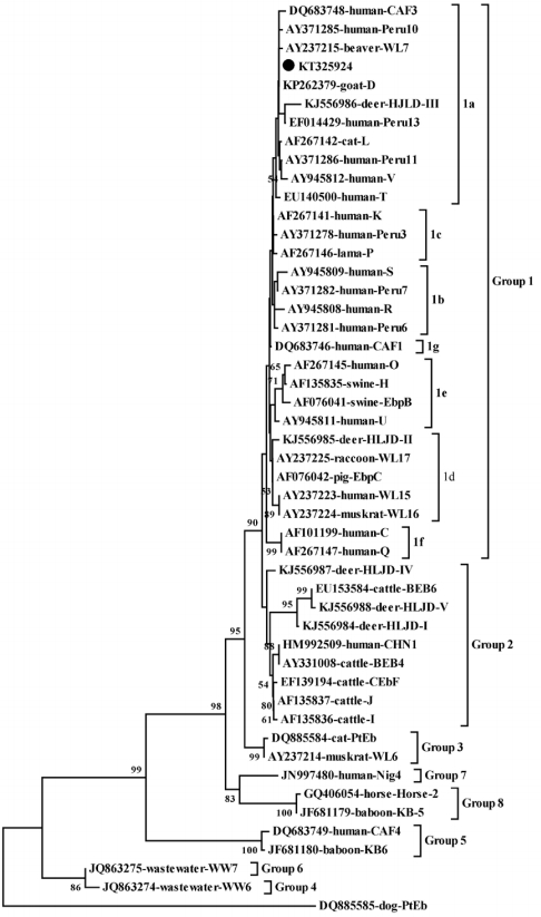
Phylogenetic analysis of Enterocytozoon bieneusi using Neighbor-Joining (NJ) method based on sequences of the internal transcribed spacer (ITS) region. The numbers at notes indicate bootstrap values (1,000 replicates, values below 50% are not shown). E. bieneusi isolate identified in the present study is indicated by solid circle.
To date, information concerning distribution of genotypes of E. bieneusi in China is scarce, especially in rabbits. In order to further improve the prevalence and distribution information, we conducted the present study. The overall prevalence of E. bieneusi infection in rabbits was 0.94%, which was considerably lower than that in other animals in northeastern China (Table 3). The lower infection rates may be related to different susceptibility of different animals, sample sizes and geo-ecological conditions, standardized management mode, and so on. Moreover, the prevalence of E. bieneusi in rabbits seemed to increase with the age in the present study, which agreed to previous studies; E. bieneusi was an opportunistic parasite and it may accumulate throughout the life time [4,5]. Moreover, probably because of similar climates in Changchun City, Jilin City, and Shenyang City, and the same management mode in these farms, the positive rates of E. bieneusi infection in rabbits among the 9 farms in northern China were not statistically different (P>0.05).

Enterocytozoon bieneusi genotypes identified in different animals in northeastern China
In the present study, only genotype D (n=3) was found in rabbits. This result was similar to a previous study conducted in Spain, in which also only genotype D was identified [6]. Genotype D not only has a wide geographical distribution but also can infect almost all host species, including humans, domestic animals, and wild animals [6]. In China, genotype D also has a broad host range. Besides some animals, e.g., golden takins (Budorcas taxicolor bedfordi) and panda in Shanxi [7,8], pet chinchillas in Anyang, Beijing, and Zhengzhou, China [9], nonhuman primates in Henan, Guangxi, Guangdong, Yunnan [10], and Sichuan [11], dairy cattle, goats, sheep, pigs, cats, and dogs in northern China [12-15]. Genotype D infection was also reported in children in Shanghai [16] and HIV patients in Henan [17], which suggest that E. bieneusi genotype D could readily transmit between humans and animals. More importantly, genotype D was also present in wastewater which may lead to outbreaks of this disease [18]. Rabbits may play an important role and cannot be ignored during this pathogen infection process in China. Moreover, sequence analysis of the ITS rRNA gene showed that the obtained sequences were identical to the reference sequence from sheep and goat isolates (accession no. KP262379) in China, which revealed that the infectious source is more likely to be excreted from sheep and goats. Therefore, we should pay serious attention to nosocomial transmission among different animals.
In view of such situation, the following practice should be performed to prevent transmission of E. bieneusi among rabbits, other animals, and humans in China: first, different kinds of animals should be managed strictly separately; second, the prevalence of E. bieneusi should be examined periodically; third, feces should be cleaned timely; fourth, parasites should be expelled regularly; fifth, humans should have a habit of wash hands before meals and after contact with animals.
In conclusion, the present study first demonstrated existence (0.94%, 4/426) of E. bieneusi infection in rabbits in China, and the infection rate was increased with age. Moreover, only genotype D was identified in rabbits which should be considered as an important potential source for E. bieneusi infection in China. Therefore, effective strategies should be taken to prevent and control of E. bieneusi infection in rabbits, other animals, and humans in China.
Acknowledgements
This work was supported by the National High-Tech R&D Program of China (863 Programs) (no. 2013AA102806, no. 2011AA10A215), National Natural Science Foundation of China (no. 31272552, no. 31272541), the Key Scientific and Technological Project of Jilin Province (no. 20140204068NY), Jilin Province Science and Technology Development Program of China (no. 20111816), and Jilin Province Quality and Safety of Agricultural Products Program by World Bank (no. 2011-Y07).
Notes
The authors declare that they have no competing interests.