Postmetacercarial changes in Echinostoma caproni maintained in a defined medium plus calf serum
Article information
Abstract
The present study examined postmetacercarial changes in the excysted metacercariae of Echinostoma caproni maintained in the defined medium Mixture 199 plus 20% calf serum for 7 days at 41℃. The gas phase was atmospheric air. Each culture was inoculated with 25 excysted metacerariae. Cultures were maintained upright in closed 15 ml plastic centrifuge tubes each containing 10 ml of medium plus 200 units of penicillin/ml and 200 µg of streptomycin/ml. By 4 days in culture, most metacercariae had voided their excretory concretions. Organisms were clumped or solitary at the bottom of the cultures. Many organisms showed flaring of the oral collar and extension of both the collar and tegumentary spines. By 4 days in culture, posterior protuberances or bumps were noted on many of the organisms and some organisms showed abnormal vesicular growths or blebs at their posterior ends. Some mortality was noted in culture by day 5, but most organisms were still alive when the cultures were terminated on day 7.
Echinstoma caproni is a good model for various studies on the biology of Echinostoma and echinostomiasis. The life cycle of this echinostome is easily maintained in the laboratory between Biomphalaria glabrata snails and Institute of Cancer Research (ICR) mice (see Fried and Huffman, 1996 for review).
Although excysted metacercariae of this echinostome have been cultivated to ovigerous adults in chick embryos (Chien and Fried, 1992), surprisingly little work has been done on the in vitro maintenance or cultivation of the metacercarial stage of this echinostome. The encysted metacercariae of this species are easy to excyst in vitro in an alkaline trypsin-bile salts medium as described in Ursone and Fried (1995); therefore, excysted metacercariae are readily available to initiate in vitro cultivation or maintenance studies.
The excysted metacercaria contains an abundant number of excretory concretions (calcareous corpuscles) in the protonephridial tubules as seen in Figure 15 in Fried and Huffman (1996). From observations of worms grown in chicks and mice, it is apparent that the excretory concretions are not present in the adults of E. caproni. Thus, they are probably voided between the metacercarial and adult stages. A purpose of the in vitro maintenance study reported herein was to note the disappearance of these concretions and also report other findings.
Metacercariae of E. caproni were removed from experimentally infected B. glabrata snails and chemically excysted in an alkaline trypsin-bile salts medium at 41℃ as described in Ursone and Fried (1995). Excysted metacercariae were transferred through three changes of sterile Locke's solution containing 200 units penicillin/ml and 200 µg streptomycin/ml. The medium used to maintain the excysted metacercariae was the defined medium Mixture 199 + 20% calf serum (Mix 199 + 20 CS). The medium was fortified with penicillin/streptomycin as described for the Locke's solution. All chemicals were purchased from Sigma (St. Louis, MO, USA). Cultures were done in 15 ml plastic centrifuge tubes containing 10 ml of the medium. The cultures were capped and maintained upright in an incubator at 41℃ for up to 7 days. The gas phase was atmospheric air. Each culture was inoculated with 25 excysted metacercariae at the start of the experiment and one culture per day was examined for 7 days.
All metacercariae examined were alive until 4 days post-inoculation. Many of the metacercariae were attached to each other in a clump at the bottom of the tubes, but some metacercariae were solitary. Metacercariae began to void their excretory concretions by day 1 so that by day 2, most metacercariae contained 4 to 8 concretions. By 2 days the collar and tegumentary spines were flared out, making them more visible than spines seen in the excysted metacercariae. By 4 days in culture, most excysted metacercariae were completely devoid of excretory concretions (Fig. 1) and some contained bumps or protuberances (Fig. 2); others contained a vacuolar bleb in the posterior aspect of the organism (Fig. 3). Some mortality was seen by day 5 with 5 of the 25 metacerariae being dead. Mortality increased to 10 of 25 dead organisms by 7 days of culture at which time the cultures were terminated. The surviving organisms looked the same at 7 days as they did at 4 days with some also showing bumps, protuberances, and blebs and all being devoid of the excretory concretions.
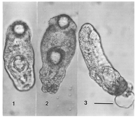
Light micrographs of excysted metacercaria of Echinostoma caproni maintained for 4 days in Mixture 199 plus 20% calf serum. Fig. 1. Intact organism showing the absence of excretory concretions. Fig. 2. Organism showing the flaring of the oral collar and protuberances in the body region posterior to the acetabulum Fig. 3. An organism showing a vacuolar bleb coming off the posterior aspect of the body. Scale bar = 90 µm and is appropriate for all 3 figures.
There has been a decline in studies on the in vitro cultivation or maintenance of digeneans. Reasons for the decline have been discussed by Irwin (1997). In brief, these studies are labor intensive and often give negative results which are not publishable. The present study used a defined medium supplemented with calf serum to examine some postmetacercarial changes in E. caproni, i.e., voidance of the excretory concretions and development of body protuberances in vitro. Future in vitro studies on this and other echinostomes may provide interesting information on the many transformational events that must occur in the development from the metacercarial to the adult stage.
In conclusion, no sexual organs appeared in 6- or 7-day-old worms. Therefore, although some somatic changes were observed, there were no indications of germinal changes in worms maintained under our in vitro conditions. Cultures were terminated at day 7 post-incubation because the culture conditions were becoming unfavorable, and many worms were dead. The appearance of the blebs, protuberance, and vesicles in the worms may be an indication of worm degeneration in an inhospitable environment. Although some digeneans have been maintained in the serum-free cultures, attempts to cultivate the excysted metacercariae of E. caproni in serum-free media were not made in our study.