INTRODUCTION
Myiasis is infection of tissues or organs of animals by fly larvae. Myiasis frequently occurs in livestock and pets in rural areas. In humans, myiasis occurs primarily in unhealthy individuals in third world countries [1]. Common infection sites are skin wounds and eyes, and nose, nasal sinuses, throat, and urogenital tract are less common sites [2]. Human myiasis may be benign and asymptomatic or may result in mild to violent disturbance, even death [3]. Only 3 other cases have been recorded in Korea; internal myiasis by Lucilia sp. (Diptera: Calliphoridae) [4], aural myiasis by Lucilia sericata [5], and nosocomial submandibular myiasis by L. sericata [6]. In this paper, we describe a nasal myiasis in a comatose patient.
CASE DESCRIPTION
A 76-year-old female, living in Cheonan-si, Chungcheongnam-do, Korea, came to the emergency room of Dankook University Hospital on 27 June 2009. She was in a state of coma due to rupture of an abdominal aortic aneurysm, and also lapsed into dyspnea. Two months prior, she was diagnosed with a large aortic aneurysm. However, she declined an aortic replacement surgery. She went to the local hospital with back pain and it was in the hospital that the aneurysm rupture occurred. She was transferred to the intensive care unit and she was put on an artificial respirator. The aneurysm was pararenal type, 8.5 × 10 cm in size. Physical examination of the patient showed abdominal distension. Surgery to repair the aortic aneurysm was performed on the day of admission.
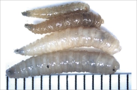
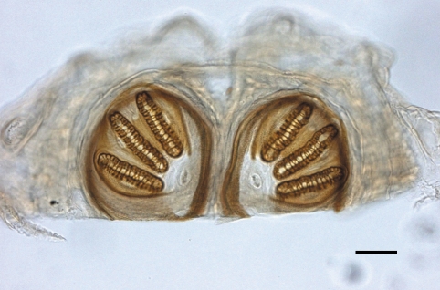
On 1 July 2009, 4 days after hospitalization, 5 fly larvae were discovered moving inside her nose (Fig. 1). They were removed endoscopically, and sent to the parasitology laboratory for analysis. Examination of the patient's nose revealed no significant pathologic changes. The larvae were white and 10.8 ± 2.6 mm × 2.0 ± 0.7 mm in size. Examination of posterior spiracles on a glass slide under light microscopy showed that they were in the family Calliphoridae, probably Lucilia sp., which is endemic in Korea (Fig. 2). They were fixed in 10% formalin and used for identification. They were identified by Lee In-Yong in Department of Environmental Medical Biology, Yonsei University College of Medicine, as the third instar larvae of Lucilia sericata. By additional endoscopic examination, no larvae were found from her. She returned from the comatose state, and did not complain any clinical signs of myiasis.
DISCUSSION
Nasal myiasis is a common disease in tropical and developing countries, but is not common in Korea. Infestations of the nose could be dangerous because of the possibility of penetration into the brain, and there is a fatality of 8% in such cases [3]. In a patient with nasal myiasis, pneumocephalus was reported as the complication [7]. The common families which cause myiasis are Sarcophagidae and Calliphorinae [8]. While Wohlfahrtia magnifica and Oestrus ovis larvae have been reported as the causative agent of nasal myiasis [3], all reported myiasis cases in Korea were by Lucilia sp. [4-6]. Lucilia sp. belongs to the family Calliphoridae and causes myiasis in humans and domestic herbivorous animals [8]. They are distributed worldwide and cause the majority of human myiasis in America, Africa, and Asia [9]. Many cases of nasal myiasis are associated with primary atrophic rhinitis [8], but it was not observed in this case.
The primary presenting symptoms of nasal myiasis include epistaxis, foul smell, pain, and sensation of foreign bodies in the nose [8]. Because the patient did not complain of any of these symptoms prior to hospitalization, the infection was likely acquired during hospitalization. In fact, the development time of Lucilia sp. is very short [10]. The first instar larvae hatch within a day, and 4 days are enough for puparium development [11]. The period between hospitalization and the discovery of larvae was 5 days, and the weather was warm when the case occurred, which may have caused faster development. Therefore, the fly probably entered the patient's nose after hospitalization, and her comatose status may have triggered its entrance.
Identification of the species prior to treatment is important because not all types of myiasis are benign [11]. In this case, the identification of the larvae was accomplished mainly by observing the posterior spiracles. It should be needed to rear larvae to adults on small pieces of meat for correct diagnosis, but this was not possible in our case due to the small number of larvae. Because the larvae can reach deep and inaccessible areas of the nose and the paranasal sinuses, the use of nasal endoscope is recommended [8]. In this case, the treatment consisted only of the removal of larvae by nasal endoscope. Considering that hospital-acquired myiasis has probably been underreported [6], control of fly populations, including the use of screens, is needed in hospitals, especially in the intensive care unit.