


Blastocystis has recently been recognized as the most prevalent eukaryotic microbe in the human gut [1], occurring worldwide in both healthy and symptomatic humans and other animals. Blastocystis is thought to be transmitted via the fecal–oral route and in cyst form [2]. Recently, PCR-based approaches using feces directly or after culture of fecal specimens have been widely used to diagnose Blastocystis infection [3]. Based on small subunit ribosomal DNA (SSU rRNA) gene analysis, this genus comprises at least 17 subtypes (STs) [4]. In human, ST1–ST4 probably account for more than 90% of carriage; the other subtypes are ST5–ST9 [5]. Although Blastocystis is of great scientific interest, neither its biology nor pathophysiology has been well-explored. Since no direct evidence indicates that Blastocystis causes diarrhea, this point also remains controversial. Only 3 Korean studies on Blastocystis have been published [6–8], all in animals. Here, we investigated the prevalence of Blastocystis and its subtypes, and the associated clinical parameters, in diarrheal and asymptomatic Korean groups.
A total of 324 stool samples were obtained from 196 diarrheal and 128 non-diarrheal subjects who underwent general checkups at Chonnam National University Hospital and Chonnam National University Hwasun Hospital, from February 2016 to October 2018. Fecal samples were collected in accordance with the guidelines of, and with the approval of, the Institutional Review Board of Chonnam National University Hospital (approval no. IRB CNUH-2015-052). We recorded age, sex, white blood cell count (WBC,×103/μl), and differential counts, red blood cell count (RBC,×106/μl), hemoglobin (Hgb, g/dl), platelet count (PLT), total protein (g/dl), albumin (g/dl), alkaline phosphatase (U/L), AST (U/L), ALT (U/L), BUN (mg/dl), creatinine (mg/dl), lactate dehydrogenase (U/L), CRP (mg/dl), and stool occult blood test data. If necessary, stool culture or multiplex PCR panel evaluation was scheduled to determine pathogens causative of diarrhea. Laboratory findings and stool culture data were analyzed by diarrhea and Blastocystis status. DNA was extracted using the Cica Geneus® DNA Prep Kit (Kanto Chemical, Tokyo, Japan) following the manufacturer’s instructions. Blastocystis was detected based on the SSU rRNA gene using the Blast-505–532 (5′–GGA GGT AGT GAC AAT AAA TC–3′) and Blast-998–1017 (5′–TGC TTT CGC ACT TGT TCA TC–3′) primers [9]. Each tube contained 8.5 μl PCR primer solution (1 μl each of Blast-505–532 and Blast-998–1017 (each 25 pmol)), 36.5 μl distilled water, and 5 μl template DNA. All PCR amplifications were performed using the TaKaRa PCR Thermal Cycler Dice Gradient (TaKaRa, Tokyo, Japan). Initial denaturation at 94°C for 3 min was followed by 30 cycles of 59°C for 30 sec and 72°C for 60 sec, and a final extension at 72°C for 5 min. The PCR products were analyzed by 1.5% (w/v) agarose gel electrophoresis with ethidium bromide staining and then sent to Macrogen (Seoul, Korea) for direct DNA sequencing. Phylogenetic analysis was performed by reference to database Blastocystis SSU rRNA genes, and a phylogenetic tree was constructed using Geneious Prime (Biomatters Ltd, Auckland, New Zealand). Phylogenetic inferences were derived using a pair-group method featuring arithmetic average clustering with 1,000 bootstrap replications. Student’s t-test was used to compare continuous variables (age and laboratory parameters). The chi-squared or Fisher’s exact test was employed to determine the distributions of categorical variables (sex and the statuses of stool occult blood, diarrhea, and Blastocystis). The likelihood-ratio chi-squared test was employed to calculate odds ratios (ORs) for Blastocystis positivity by subtype. All statistical analyses were performed using SPSS ver. 25.0 software (SPSS Inc., Chicago, Illinois, USA). A P-value <0.05 was considered to indicate significance.
Of the 324 samples, 29 (9.0%) including 6 diarrheal and 23 non-diarrheal samples were positive for Blastocystis, with a significant difference between the 2 groups (P<0.001) (Table 1). Thus, Blastocystis may not necessarily cause diarrhea. Blastocystis has previously been found in both symptomatic and asymptomatic patients [10–12]. It has been suggested that intra-subtype variation at the SSU rRNA gene level might affect the presenting symptoms of patients with identical Blastocystis subtypes [13]. We found subtypes ST1, ST2, ST3, and multiple infections in 3 (50.0%), 0 (0.0%), 2 (33.3%), and 1 (16.7%) patient in the diarrheal group and in 4 (17.4%), 1 (4.3%), 18 (78.3%), and 0 (0.0%) patient the non-diarrheal group. Of the 3 subtypes, ST3 was more common in the non-diarrheal group (78.3 vs. 33.3%, P=0.0341). ST1 seemed to be frequently found in diarrheal group compared to non-diarrheal group, but no statistical significance (diarrheal group vs. non-diarrheal group, 3/6 vs. 4/23, P=0.0964). Further study may be necessary to determine potential correlation of certain subtypes and symptoms. We did not detect ST4, in agreement with previous studies suggesting that this ST was rare in Asia and the Middle East [14,15]. When we analyzed the distribution of STs, a total of 26 sequenced samples (GenBank accession no. MT186203-MT186218 enrolled in this study) were closely related to the sequences of each known human STs (Fig. 1). All but 2 ST3 sequences clustered together with sequences from the other countries such as Germany (MK801366, MK801409), Singapore (KX618192), Poland (MN918265), Mexico (MK874780, KU147402), South Korea (MT093452), Iran (MH049544, LC414153) and Thailand (JX305884), respectively. Notably, 2 ST3 sequences (MT186204, MT186206) from the diarrheal group were somewhat distant from the others; ST3 thus exhibited intra-genetic variation. All sequences lay distant from those of ST4 (KX351997, KP284173), ST5 (MT094303, KM438216), ST6 (MG011651, KP284174), and ST7 (JN003686), respectively. We also analyzed several laboratory parameters by Blastocystis presence or symptoms. The white and red blood cell counts, hemoglobin level, percentages of lymphocytes, monocytes, eosinophils, and basophils, and the levels of total protein, albumin, BUN, lactate dehydrogenase, gamma-glutamyl transpeptidase, and C-reactive protein, and the rate of positive results on stool occult blood tests differed significantly between the diarrheal and non-diarrheal groups. However, only the creatinine level differed significantly between the Blastocystis-positive and -negative groups (3.6 vs. 1.1 mg/dl, P=0.001) (Table 2). Of the 196 diarrheal patients, 54 exhibited other causative pathogens (Clostroides difficile with or without other pathogens (23), Campylobacter spp. with or without other pathogens (15), Salmonella spp. (9), Citrobacter freundii (1), Cryptosporidium spp. (1), Enterococcus faecalis (1), ETEC LT/ST, STEC stx1/sb2 (1), Norovirus GI/GII (1), Giardia spp. (1) and Shigella spp. (1)). Of the 29 Blastocystis-positive patients, all but 2 (one with Campylobacter sp. and one with Giardia sp.) were negative for other possibly causative pathogens. Overall, our data support the suggestion that Blastocystis may not be pathogenic.
This is the first report of Blastocystis infections in Koreans. Blastocystis was found in both the diarrheal and non-diarrheal groups, but the subtype prevalence differed between the groups. Any role played by Blastocystis in human health and disease should be explored further.
Fig. 1
Phylogenetic tree of 26 sequences of Blastocystis SSU rRNA gene compared to database Blastocystis SSU rRNA genes. All of sequences of this study were enrolled to GenBank database (GenBank accession no. MT186203-MT186218), which were indicated in bold.
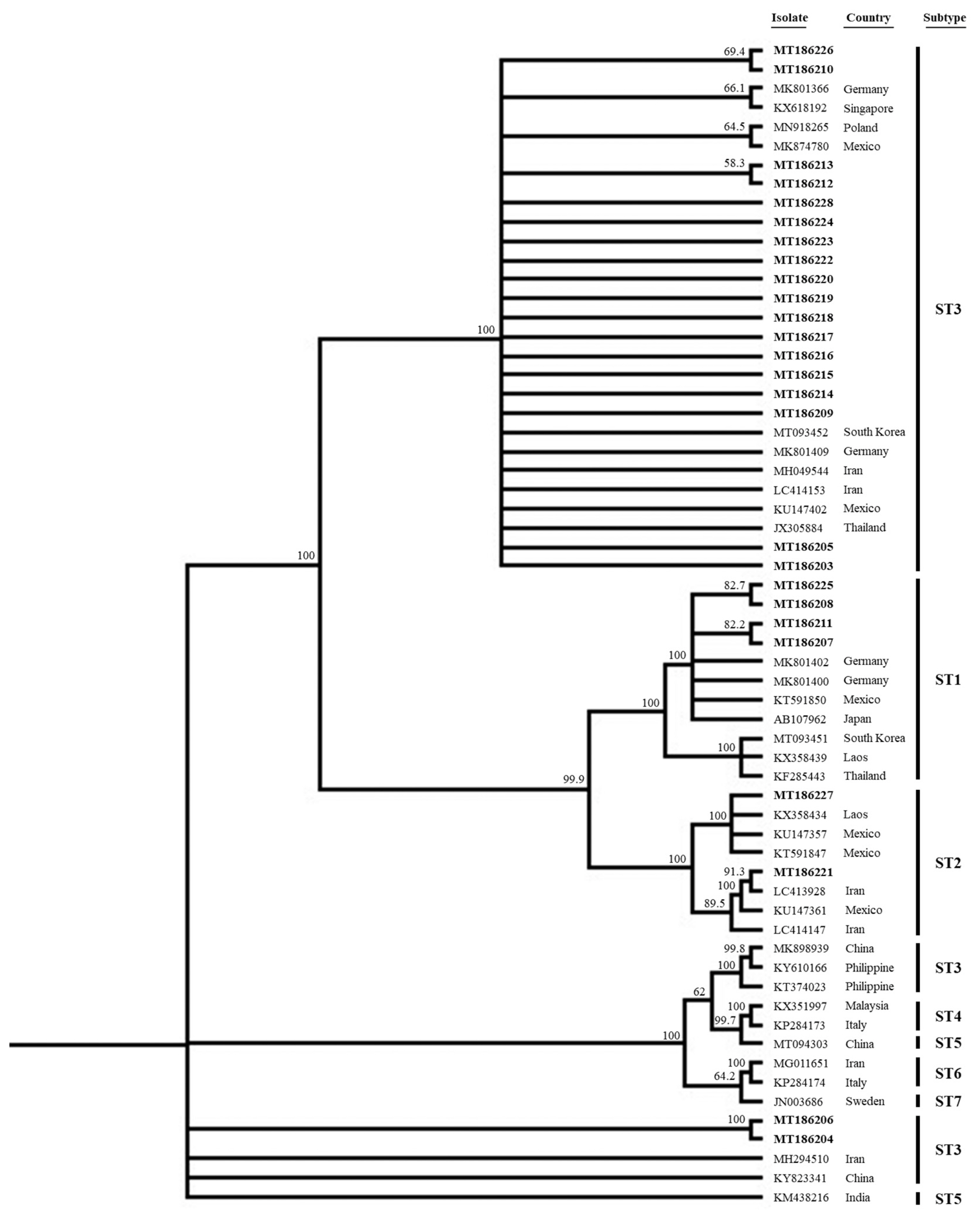
Table 1
Characteristic of laboratory findings according to the diarrheal symptoms
| Clinical parametersa | Laboratory characteristics of | P-value | |
|---|---|---|---|
| Diarrheal group (n=196) | Healthy control (n=128) | ||
| Age (yr) | 60.8±17.78 | 62.6±11.37 | 0.318 |
| WBC count (×103/μl) | 9±5.61 | 5.6±1.43 | <0.001 |
| RBC count (×106/μl) | 3.6±0.66 | 4.8±0.43 | <0.001 |
| Hemoglobin (g/dl) | 10.9±2.22 | 14.1±1.33 | <0.001 |
| Platelet count (×103/μl) | 219.6±136.82 | 252.3±54.3 | 0.13 |
| Lymphocytes (%) | 15.8±11.27 | 34.9±8.01 | <0.001 |
| Monocytes (%) | 9.3±7.54 | 6.8±2.18 | 0.034 |
| Neutrophils, (%) | 73.1±14.94 | 66.1±73.8 | 0.224 |
| Eosinophils (%) | 1.9±2.21 | 2.8±2.69 | 0.015 |
| Basophils (%) | 0.4±0.38 | 0.7±0.27 | <0.001 |
| Total protein (g/dL) | 6±0.99 | 7.5±0.48 | <0.001 |
| Albumin (g/dL) | 3.3±0.75 | 4.4±0.3 | <0.001 |
| ALP (U/L) | 114.4±134.06 | 75.7±22.59 | 0.058 |
| AST (U/L) | 36.1±38.3 | 29±12.01 | 0.067 |
| ALT (U/L) | 28.4±29.73 | 27.7±18.33 | 0.829 |
| Total Bilirubin (mg/dL) | 1.2±2.11 | 0.8±0.33 | 0.348 |
| BUN (mg/dl) | 17.6±14.35 | 12.1±4.89 | 0.011 |
| Creatinine (mg/dl) | 1.2±3.36 | 1.3±3.51 | 0.812 |
| Lactate dehydrogenase (U/L) | 618.9±450.65 | 141.5±94.76 | <0.001 |
| r-GTP (IU/L) | 109±147.21 | 31.5±28.56 | <0.001 |
| CRP (mg/dl) | 7.8±7.69 | 0.1±0.13 | <0.001 |
| Sex (male) | 116/196 (59.2) | 54/128 (42.2) | 0.003 |
| Presence of abdominal pain No. (%) | 92/196 (46.9) | 4/128 (3.1) | <0.001 |
| Positive for stool occult blood No. (%) | 9/23 (39.1) | 0/86 (0.0) | <0.001 |
| Positive for Blastocystis, No. (%) | 6/196 (3.1) | 23/128 (18.0) | <0.001 |
| Blastocystis subtype No. (%) | |||
| ST1 | 3/6 (50.0) | 4/23 (17.4) | 0.0964 |
| ST2 | 0/6 (0.0) | 1/23 (4.3) | |
| ST3 | 2/6 (33.3) | 18/23 (78.3) | 0.0341 |
| Multi band | 1/6 (16.7) | 0/23 (0.0) | |
Table 2
Characteristic of laboratory findings according to the presence of Blastocystis
| Clinical parametersa | Laboratory characteristics | P-value | |
|---|---|---|---|
| Blastocystis-negative (n=295) | Blastocystis-positive (n=29) | ||
| Age (yr) | 61.4±15.94 | 62.6±11.48 | 0.692 |
| WBC count (×103/μl) | 8.5±5.29 | 6.5±4.75 | 0.234 |
| RBC count (×106/μl) | 3.8±0.76 | 4.1±0.9 | 0.301 |
| Hemoglobin (g/dl) | 11.9±2.48 | 12.7±2.2 | 0.147 |
| Platelet count (×103/μl) | 226.6±128.34 | 197.7±82.64 | 0.482 |
| Lymphocytes (%) | 19.1±13.11 | 23.9±11.13 | 0.254 |
| Monocytes (%) | 8.8±6.99 | 8.9±5.8 | 0.965 |
| Neutrophils (%) | 72.1±34.9 | 64.4±13.94 | 0.488 |
| Eosinophils (%) | 2±2.35 | 2.2±2.1 | 0.808 |
| Basophils (%) | 0.4±0.39 | 0.5±0.38 | 0.495 |
| Total protein (g/dl) | 6.3±1.09 | 6.5±1.07 | 0.606 |
| Albumin (g/dl) | 3.5±0.82 | 3.9±0.56 | 0.083 |
| ALP (U/L) | 107.5±121.95 | 64.9±14.12 | 0.357 |
| AST (U/L) | 34.3±32.85 | 24.8±7.42 | 0.177 |
| ALT (U/L) | 28.5±27.09 | 23.5±10.74 | 0.387 |
| Total Bilirubin (mg/dl) | 1.1±1.93 | 0.7±0.2 | 0.515 |
| BUN (mg/dl) | 16.7±13.25 | 13±12.89 | 0.299 |
| Creatinine (mg/dl) | 1.1±2.82 | 3.6±7.37 | 0.001 |
| Lactate dehydrogenase (U/L) | 499.6±446.27 | 225.9±199.58 | 0.109 |
| r-GTP (IU/L) | 70.8±112.65 | 25.6±17.51 | 0.137 |
| CRP (mg/dl) | 6.8±7.69 | 5.5±5.94 | 0.613 |
| Sex male No. (%) | 159/295 (53.9) | 11/29 (37.9%) | 0.12 |
| Presence of abdominal pain No. (%) | 93/295 (31.5) | 3/29 (10.3%) | 0.018 |
| Positive for stool occult blood No. (%) | 9/95 (9.5) | 0/14 (0.0%) | 0.601 |
| Positive for Diarrhea No. (%) | 190/295 (64.4) | 6/29 (20.7%) | < 0.001 |
| Other pathogens No. (%) | 52/295 (17.6)b | 2/29 (6.9%)c | |
a Clinical parameters, except for Sex, Stool occult blood, Diarrhea, Other pathogens, were expressed with Mean±SD.


