INTRODUCTION
Echinostoma hortense is a member of the Echinostomatidae family, and is characterized by a large elongated body, a head crown with collar spines, a large oral sucker, tandem testes in the anterior body, and rich vitelline glands in the posterior body. The fluke inhabits the small intestine and prefers the upper region. Human echinostomatid infections, about 120 cases, have been reported in Korea. The echinostomatid species with a parasitologic history in Korean are Echinostoma hortense, E. cinetorchis, Echinochasmus japonicus and Acanthoparypium tyosenense (Seo et al., 1983, 1985; Ryang et al., 1985; Chai et al., 1994, 2001). Of these E. hortense is a dominant species in Korea. The mountainous villages of inland Korea represent its endemic foci (Lee et al., 1988). Recently human E. hortense infections were diagnosed by gastroduodenal endoscopy in Korea (Chai et al., 1994; Lee and Hong, 2002; Cho et al., 2003). Here we describe a case of infection by E. hortense in the duodenal bulb, which was diagnosed by upper endoscopy.
CASE RECORD
A Korean woman, 55-year-old, visited a local clinic with complaints of upper abdominal pains and discomfort on October 1999. Her symptoms had persisted for 2 weeks at the time of her visit. She lived in Ulsan-city, Gyeongsangnam-do, Korea. During gastroduodenal endoscopy, two motile flukes were found attached to the duodenal bulb. The flukes were slender, elongate, about 5 mm in length, with a red spot at anterior end (Fig. 1). The flukes were removed from the duodenal bulb with endoscopic forceps. The patient was treated with single dose of praziquantel, 10 mg/kg. After treatment, the abdominal symptoms were subsided.
She had worked in a small restaurant, and had eaten raw frog meat several times about two months before onset of her symptoms. She denied ever eating freshwater fish in raw. Laboratory data were not available. A stool examination revealed six echinostome eggs, but none were found in family members. The eggs were bright yellow, elliptical, thin shelled with a shallow operculum, and measured 114.1 (111.2-118.6) µm long and 76.7 (74.1-79.0) µm wide (Fig. 2).
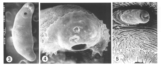
The flukes were processed for parasitological identification by light and scanning electron microscopic studies. The anterior of the fluke was bent ventrally at the acetabulum, and the surface was wrinkled transversely over the whole body. The body surface was covered with cobblestone-like cytoplasmic processes. Tegumental spines, scale-shaped with a broad base, were densely packed on the anterior surface of the body and then reduced in density and size on moving posteriorly (Fig. 3). The anterior end bulged into a head crown around the oral sucker. The head crown was armed with uninterrupted 27 collar spines, of which the 4 end-group spines were present at the both ends of the the head crown (Fig. 4). The outer surface of oral sucker was smooth and encircled with ciliated sensory (Type I) papillae arranged in 2-3 rows. Unciliated sensory (Type II) papillae, dome-like round structures, appeared on posterior half of the oral sucker lip. The acetabulum, wide-open, was aspinous with velvety cytoplasmic processes, and surrounded with Type I papillae. The cirrus erected from the genital opening revealed a velvety tegumental surface, and dense Type I sensory papillae, as compared with other parts of the body surface (Fig. 5).
DISCUSSION
The number of collar and end-group spines is species-identifying morphological characteristic of echinostomatid flukes. The echinostomes retrieved from the present patient under scanning electron microscopy revealed 27 and 4 spines, which is a species-characteristic of E. hortense. Surface ultrastructures, such as the arrangement of Type I sensory papillae on the oral sucker and around the acetabulum, and scale-shaped tegumental spines distributed at decreasing density posteriorly are consistent with previous descriptions of E. hortense (Lee et al., 1986b). After referring to surface ultrastructural characteristics and egg morphology (Seo et al., 1983), the isolated flukes were identified as E. hortense.
Adult E. hortense inhabits the upper part of the small intestine in mammalian final hosts. The flukes produce ulcerative lesions in intestinal mucosa, villous atrophy characterized by blunting, villous fusion, and loss of villous tips, and crypt hyperplasia. In the stroma of villi were found inflammatory cell infiltration, vascular congestion, and edema (Lee et al., 1990). The collar spines in the head crown of E. hortense were stout and damaged the intestinal mucosa. Ulcerative lesions produced by adult E. hortense in the duodenal bulb were previously observed by upper endoscopy (Chai et al., 1994). In consideration of the pathologic findings, the symptoms experienced by the present patient, i.e., epigastric pain and abdominal discomfort, might be provoked by the abrasive action of E. hortense. The frequent symptoms reported by infected individuals, though not pathogen-specific, are abdominal pain, cramps, anorexia, postprandial burning, flatulence, and diarrhea (Lee et al., 1986a; Chai et al., 1994; Lee and Hong, 2002). It has also been suggested that chronic and severe infection may cause intestinal villous atrophy and lead to manifestations of malabsorption.
Freshwater fish have been recorded as infection sources of echinostomiasis hortense, most frequently loaches in Korea, followed by frogs (Chai et al., 1985; Ryang et al., 1985; Cho et al., 2003). Frog tadpoles become infected with metacercariae of echinostomes and metamorphose to adult frogs with the metacercariae in their tissues, predominantly in muscles. Metacercariae were previously found mainly in the dorsal pharyngeal wall, body cavity, and mesentery of Rana nigromaculata (Rim et al., 1982). People may become infected by eating inadequately cooked frog meat, as occurred in the present case. The reservoir hosts have been recognized to play a significant role in an ecosystem of digenean trematodes. They initiate the trematode's life cycle by supplying trematode eggs to the first intermediate molluscan hosts. Of reservoir hosts, rats and dogs are reported to show a high prevalence of E. hortense infection in Korea (Seo et al., 1981). This human case reports that frogs are a source of human echinostome infections in Korea.